Neurofibromatosis

Neurofibromatosis (NF) is a condition that causes tumors to grow on nerve tissue, producing skin and bone abnormalities.

NF is often diagnosed in childhood, occasionally in infancy (in children with severe cases), but usually around 3-16 years of age. Effects of the disease vary widely — some children live almost unaffected by the condition; rarely, others might be severely disabled.

There's no specific cure for NF, but tumors usually can be removed and related complications treated. Because learning disabilities occur in about half the children with NF, some might need extra help in the classroom.

About Neurofibromatosis

Neurofibromatosis is a neurocutaneous syndrome passed down through the parents' genes, and it affects the brain, spinal cord, nerves, skin, and other systems in the body.

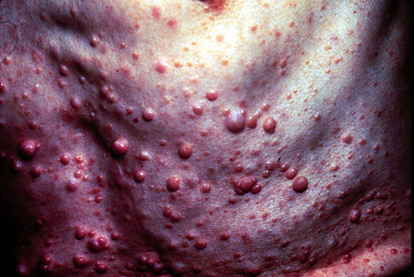
Neurofibromatosis is defined by tumors, called neurofibromas, that grow along nerves in the body, or on or under the skin. As the tumors increase in size, they can press on vital areas of the body, causing problems in the way the body functions.

Neurofibromas often first appear in childhood, especially during puberty. The first noticeable sign is almost always the presence of brown cafĂ© au lait spots. These distinctive spots don't hurt or itch and never progress to anything more serious than spots. They can be found anywhere on the body, though not usually on the face. Tiny ones — freckles — may be seen under the arms or in the groin area.

Many neurofibromas can be removed. Although usually benign (noncancerous), an estimated 3%-5% become cancerous.

Of the two types of neurofibromatosis — NF1 and NF2 — NF1 is more common, occurring in 1 of every 4,000 births and affecting an estimated 100,000 Americans. It is also known as von Recklinghausen disease.

NF2 is characterized by the presence of bilateral acoustic neurofibroma-like tumors and is rarer, seen in 1 in 50,000 births. People with NF2 usually develop benign tumors on the nerves in their ears, causing hearing loss, eventual deafness, and problems with balance.

The severity of both types of neurofibromatosis varies greatly. In families where more than one person has NF, it can present with different physical signs and complications for each person. At diagnosis, it isn't possible to know right away whether a case will be mild or lead to severe complications.

Causes of NF

Both types of neurofibromatosis are autosomal dominant genetic disorders, which means an affected person has 1 chance in 2 of passing it on with each pregnancy. Neurofibromatosis also can be the result of a spontaneous change (mutation) in the genetic material of the sperm or egg at conception in families with no previous history of NF. About half of cases are inherited, and the other half are due to spontaneous genetic mutation.

NF 1 and NF 2 are each related to changes in separate genes. The NF1 gene is located on chromosome 17, and NF2 has been traced to chromosome 22. These findings are important because they may eventually lead to the development of a blood test or other genetic test to find out if a relative has NF.

Signs and Symptoms

NF1 is sometimes diagnosed in younger children, especially those with more severe forms of the disorder. One key to early diagnosis of mild NF is the appearance of café-au-lait spots on the skin.

Many people who do not have NF have a few cafĂ©-au-lait spots. But if a young child has five or more, at least ½ inch in size (roughly the size of a dime), a doctor will look for other clues that may indicate NF, including neurofibromas — tumors on, under, or hanging off the skin — and Lisch nodules, tiny, noncancerous tumors on the iris (the colored part of the eye). Lisch nodules are of no clinical significance except that they help confirm a diagnosis of NF.

Neurofibromas often become evident on various parts of the body, beginning at the arms, around 10 years of age. A child may also develop freckling in the folds of the skin of the armpit or groin or on other parts of the body where the skin creases.

Abnormalities of the skeleton, such as the thinning or overgrowth of the bones in the arms or lower leg, curvature of the spine (scoliosis), and other bone deformities also may be features of NF1.

NF2 is usually not diagnosed until a child is older. Hearing loss in the late teens and early twenties is often among the first symptoms of the disorder, and is caused by tumors growing on the auditory nerves (which carry electrical impulses from the inner ear to the brain, allowing us to hear) on one or both sides.

Other symptoms of NF2 include continuous ringing in the ears, headache, facial pain or weakness, and feeling unsteady or off balance.

Diagnosis and Treatment

Neurofibromatosis is usually diagnosed based on a combination of findings. A child must have at least two of the following signs to be diagnosed with NF1:

  • cafĂ©-au-lait spots of a certain number, size, and location
  • the appearance of two or more neurofibromas (often resembling pea-sized bumps on the skin)
  • Lisch nodules on the irises
  • an optic glioma (tumor along the main nerve of the eye that is responsible for sight)
  • certain skeletal abnormalities
  • a family member with NF1
  • freckling under the arms or in the groin

Tests like magnetic resonance imaging (MRI) and X-rays may be used to screen for tumors or evidence of skeletal problems. A child's head circumference will be measured, as kids with symptoms of NF can have a circumference that's larger than normal for their age. Blood pressure will be monitored. Doctors also take a detailed personal history, looking for signs of learning difficulties.

To diagnose NF2, doctors will check for any evidence of hearing loss. They'll order audiometry (hearing tests) as well as imaging tests to look for tumors in the nerves of the ears, spinal cord, or brain. They'll also determine if there's a family history of NF2.

Genetic testing is now available for people with a family history of either NF1 or NF2, though such testing is still not 100% sensitive. Amniocentesis or chorionic villus sampling can sometimes determine if an unborn child has the condition.

Treatment for NF1 includes removal of the neurofibromas for cosmetic purposes, treating the complications (see below), and getting intervention for children with learning disabilities. Kids will be referred to appropriate medical specialists to monitor and treat complications, which may include:

  • seizures (up to 40% of children with NF1 have them)
  • high blood pressure
  • scoliosis
  • speech impairment
  • optic nerve tumors (which can cause vision problems leading to blindness)
  • early or delayed onset of puberty

Rarely, neurofibromas can become cancerous (3%-5% of cases). In these occurrences, surgery, chemotherapy, or radiation may be necessary.

With NF2, surgeons will likely need to remove the auditory nerve tumors, which may cause deafness afterward. When parts of the auditory nerve are removed, hearing aids won't work.

In 2000, the U.S. Food and Drug Administration (FDA) approved an auditory brainstem implant for people with NF2 who have lost their hearing. This device transmits sound signals directly to the brain, enabling the person to hear certain sounds and speech.

Currently, researchers are conducting trials with medications in the hopes they'll be able to offer more treatment options.

Caring for Your Child

The first noticeable sign of neurofibromatosis usually is the presence of multiple café-au-lait spots. If your child has several of these spots, ask your doctor to do a thorough examination; he or she may need to screen your child for other signs of NF.

If your child has already been diagnosed with NF and you notice that a growing tumor is beginning to cause a problem, tell your doctor immediately.

One of the most important things you can do is get early intervention if your child has learning disabilities. It also helps to seek out support groups that can provide your family with practical advice and encouragement.

Remember, most people (about 60%) diagnosed with NF1 have only relatively mild signs of the disorder, like café-au-lait spots and a few neurofibromas on the surface of the skin, which require little or no treatment.

Kids diagnosed with mild NF who remain fairly healthy into early adulthood are less likely to develop more serious complications later in life. Kids diagnosed with more serious forms often have correctable complications and with appropriate help and support can lead happy and productive lives.

Neurocutaneous Syndromes

Neurocutaneous syndromes are disorders that lead to abnormal growth of tumors in various parts of the body. They're caused by the abnormal development of cells in an embryo and characterized by the presence of tumors in various parts of the body (including the nervous system) and by certain differences in the skin.

While some can be diagnosed at birth, others don't produce symptoms until later in life. Although neurocutaneous syndromes cannot be cured, treatments can help manage symptoms and any health problems that occur.

Types of Neurocutaneous Syndromes

Common neurocutaneous syndromes that affect kids include:

  • neurofibromatosis, types 1 and 2 (NF1 and NF2)
  • Sturge-Weber syndrome
  • tuberous sclerosis (TS)
  • ataxia-telangiectasia (A-T)
  • von Hippel-Lindau disease (VHL)

Symptoms vary widely from condition to condition, and they affect different kids in different ways. Often, the full effects of these diseases — even if detected at birth — do not emerge until the child grows up. The educational, social, and physical problems that the conditions cause must be managed throughout a child's life.

Neurofibromatosis

Neurofibromatosis is one of the most common neurocutaneous syndromes. It can cause tumors to grow on nerve cells, producing skin changes, bone deformities, eye problems, and other complications, particularly in the brain.

Neurofibromatosis is usually inherited, but up to half of cases occur because of spontaneous changes (mutations) within a person's genes. Once a mutation has taken place, the changed (mutant) gene can then be passed on to succeeding generations. The child of a parent with neurofibromatosis has a 50% chance of inheriting the disorder.

The two different forms of this disorder are neurofibromatosis type 1 (NF1) and neurofibromatosis type 2 (NF2). NF1 accounts for approximately 90% of all cases.

Neurofibromatosis Type 1

NF1 (also known as von Recklinghausen disease) occurs in about 1 in 4,000 babies born in the United States. To diagnose NF1, doctors take a thorough medical and family history because children with NF1 often have a parent with the disease.

The classic sign of NF1 are skin pigment findings known as "café-au-lait" spots. These light brown or coffee-colored patches may be present at birth and can look like freckles at first. They often increase in size and number during the first few years of life. A child diagnosed with NF1 will usually have at least six café-au-lait spots that are larger than freckles. The spots are flat, don't itch or hurt, and do not turn into anything more serious.

Another common sign is the presence of Lisch nodules, tiny, benign (noncancerous) tumors found on the iris of the eye. In some cases, tumors can develop along the optic nerves and affect vision. During puberty, benign tumors called neurofibromas develop on or under the skin or along the nerves of the body. Bone deformities also may develop.

NF1 treatment focuses on managing the symptoms. A child with complications involving the eye, nervous system, spine, or bones will be referred to an appropriate specialist for treatment. In cases where these neurofibromas are causing chronic pain, growing into vital body organs, or causing infections, the growths can be removed through surgery.

Children with NF1 also have a high prevalence of seizures, learning disabilities, attention deficit disorder (ADHD), and speech problems. Therapy and specialists can help manage those symptoms.

Neurofibromatosis Type 2

Neurofibromatosis type 2 is less common, occurring in about 1 in 40,000 births. Kids who have it usually develop tumors on the auditory nerves (the nerves leading to the ear), but not until adolescence or as young adults.

NF2 symptoms, which appear in the teen years or early twenties, can include hearing loss, ringing of the ears, and problems with balance. Different treatment options can help manage these problems.

Tuberous Sclerosis

Tuberous sclerosis, or TS, causes benign growths called tubers to form on different body organs, including the brain, eyes, kidneys, heart, skin, and lungs. It occurs in approximately 1 in 6,000 births, and the child of a parent with TS has a 50% chance of having it.

TS is often first recognized when a child has seizures or shows developmental delays. The severity of TS symptoms vary greatly among kids, ranging from mild skin abnormalities to mental retardation or kidney failure.

Treatment usually includes medication to prevent seizures, treatments to address skin problems, surgery to remove tumors, and the management of high blood pressure caused by kidney disease.

Sturge-Weber Syndrome

Sturge-Weber syndrome is a rare condition caused by a spontaneous genetic mutation that affects the skin and the brain. What prompts the mutation is unknown and so far no genetic factors have been found. It is not passed down by parents who carry the disease. Because it frequently goes undiagnosed, it's difficult to estimate how many people are affected.

Each case of Sturge-Weber is unique and symptoms vary widely. The most visible marker is a facial birthmark or "port-wine stain" that is present at birth and usually covers at least one upper eyelid and the forehead.

Sturge-Weber syndrome can lead to neurological problems, including unusual blood vessel growths on the brain called angiomas. These often cause seizures that begin before the first birthday and can worsen with age. A child also may experience convulsions on the side of the body that's opposite from the port-wine stain.

About 30% of patients also develop glaucoma (increased pressure inside the eye that impairs vision), typically in the eye that is affected by the port-wine stain. That eye also might be enlarged (a condition called buphthalmos). Some kids who have this condition also experience strokes.

Treatments, medications, and surgery can help a child cope with the health problems associated with the disease.

Children as young as 1 month old who have Sturge-Weber can undergo laser treatment to reduce or remove port-wine stains. Anticonvulsant medication may be used to control seizures, and surgery can control glaucoma and vision problems.

Ataxia Telangiectasia

Ataxia telangiectasia (A-T) is a progressive degenerative disease involving many major body systems. It is a recessive genetic disease, meaning that both parents carry the gene that could combine to cause A-T in their children but do not have the disease themselves. Two parents who carry the mutated gene have a 25% chance of having a child affected by A-T.

A-T is usually noticed in the second year of life as a child develops problems with balance and slurred speech caused by ataxia (lack of muscle control). The ataxia occurs because the cerebellum, the part of the brain that controls muscle movement, is degenerating. Eventually, the lack of muscle control becomes severe enough for the child to require a wheelchair.

Another symptom of A-T is the appearance of tiny, red, spiderlike veins in the corners of the eyes or on the ears and cheeks when exposed to sunlight. These veins, known as telangiectasias, are harmless.

About 70% of children with A-T also have immune system problems that make them more susceptible to chronic upper respiratory infections, lung infections, and pneumonia. They're also very susceptible to developing certain cancers, such as leukemia and lymphoma.

Currently, there is no treatment for A-T and no way to stop its progression. But treatment can help kids manage symptoms. Physical therapy and occupational therapy may help maintain flexibility, and speech therapy can help address slurring and other speech problems. Special medications may be given to help enhance weakened immune systems.

Von Hippel-Lindau Disease

Von Hippel-Lindau disease (VHL) is a genetic disorder involving the abnormal growth of blood vessels. It usually affects certain areas, such as the brain and other parts of the central nervous system, the retina of the eye, the adrenal glands, the kidneys, or the pancreas. Its prevalence is unknown, but the child of a parent who carries the gene that causes VHL has a 50% chance of having the disorder.

Blood vessels usually grow like branches on a tree, but in kids with VHL, they form small tumors called angiomas. Doctors carefully monitor angiomas because, depending on where they are located, they can cause other medical problems. For example, angiomas on the retina of the eye may lead to vision loss.

Symptoms usually appear between 10 and 30 years of age. VHL is diagnosed through magnetic resonance imaging (MRI) or a computerized tomography (CT) scan. A thorough physical examination and blood tests are also performed.

Symptoms depend on the size and location of the angiomas, and can include headaches, balance problems, dizziness, weakness, vision problems, and high blood pressure. Fluid-filled cysts or tumors (benign or cancerous) may develop around the angiomas, worsening these symptoms. People with this disorder have a higher risk of developing cancer, especially kidney cancer.

VHL treatment will depend on the size and location of the angiomas. The goal is to treat the tumors while they're small and before they put pressure on any of the major organs, such as the brain or spine. Surgery may be required to remove the tumors before they create severe problems.

The prognosis for VHL patients depends on the location of the tumors and the complications they cause. Fortunately, early detection and treatment can improve a child's treatment outcome.

Caring for Your Child

The illnesses associated with neurocutaneous syndromes can place enormous stress and emotional burdens on you and your child, and it is easy to feel overwhelmed. Early intervention is important to helping your child achieve the best quality of life possible.

The focus of treatment is to prevent or minimize complications and maximize the child's strengths. Keep in mind the following tips:

  • Positive reinforcement can strengthen your child's self-esteem and foster a sense of independence. Let your child find out what he or she is capable of, especially regarding daily living skills.
  • Support groups can be extremely beneficial, so seek out local chapters that address your child's particular illness. They provide a supportive social environment, and are a great way to share knowledge and resources.
  • Psychotherapy or other supportive treatments can boost your child's self-esteem and coping skills, so ask the treatment team for referrals. Therapy also can help other family members deal with the stress involved in caring for a child with a chronic illness or disability.
  • Physical, occupational, or speech therapy can help your child improve some of the developmental delays caused by the specific illness.
  • Check with your local hospital or university for seminars or informational classes about neurocutaneous syndromes. Education can help you be a valuable resource in your child's long-term treatment.

Many medical professionals might care for your child during diagnosis and treatment. These professionals can include a family practitioner, pediatrician, neurologist, neurosurgeon, orthopedic surgeon, oncologist, geneticist, and ophthalmologist. A genetic counselor also can provide information about genetic testing and the risk of passing the disease on to another child.

Remember that although each of these conditions are challenging, supportive therapies and treatments can help both you and your child.

Muscular Dystrophy

Muscular dystrophy (MD) is a genetic disorder that gradually weakens the body's muscles. It's caused by incorrect or missing genetic information that prevents the body from making the proteins needed to build and maintain healthy muscles.

A child who is diagnosed with MD gradually loses the ability to do things like walk, sit upright, breathe easily, and move the arms and hands. This increasing weakness can lead to other health problems.

There are several major forms of muscular dystrophy, which can affect the muscles to varying degrees. In some cases, MD starts causing muscle problems in infancy; in others, symptoms don't appear until adulthood.

There is no cure for MD, but researchers are quickly learning more about how to prevent and treat it. Doctors are also working on improving muscle and joint function and slowing muscle deterioration so that those with MD can live as actively and independently as possible.

First Symptoms

Many kids with muscular dystrophy follow a normal pattern of development during their first few years of life.

But in time common symptoms begin to appear. A child who has MD may start to stumble, waddle, have difficulty going up stairs, and toe walk (walk on the toes without the heels hitting the floor). A child may start to struggle to get up from a sitting position or have a hard time pushing things, like a wagon or a tricycle.

Kids with MD often develop enlarged calf muscles (called calf pseudohypertrophy) as muscle tissue is destroyed and replaced by fat.

Diagnosis

When first suspecting that a child has muscular dystrophy, a doctor will do a physical exam, take a family history, and ask about any problems — particularly those affecting the muscles — that the child might be having.

In addition, the doctor may perform tests to determine what type of MD is involved and to rule out other diseases that could cause the problem. These might include a blood test to measure levels of serum creatine kinase, an enzyme that's released into the bloodstream when muscle fibers are deteriorating. Elevated levels indicate that something is causing muscle damage.

The doctor also may do a blood test to check the DNA for gene abnormalities or a muscle biopsy to examine a muscle tissue sample for patterns of deterioration and abnormal levels of dystrophin, a protein that helps muscle cells keep their shape and length. Without dystrophin, the muscles break down.

Types of Muscular Dystrophy

The different types of muscular dystrophy affect different sets of muscles and result in different degrees of muscle weakness.

Duchenne muscular dystrophy is the most common and the most severe form of MD. It affects about 1 out of every 3,500 boys. (Girls can carry the gene that causes the disease, but they usually have no symptoms.) This form occurs because of a problem with the gene that makes dystrophin. Without this protein, the muscles break down and a child becomes weaker.

In cases of Duchenne muscular dystrophy, symptoms usually begin to appear around age 5, as the pelvic muscles begin to weaken. Most kids with this form need to use a wheelchair by age 12. Over time, their muscles weaken in the shoulders, back, arms, and legs.

Eventually, the respiratory muscles are affected, and a ventilator is required to assist breathing. Kids who have Duchenne MD typically have a life span of about 20 years.

Although most kids with Duchenne muscular dystrophy have average intelligence, about one-third of them experience learning disabilities and a small number have mental retardation.

While the incidence of Duchenne is known, it's unclear how common other forms of MD are because the symptoms can vary so widely between individuals. In fact, in some people the symptoms are so mild that the disease goes undiagnosed.

Becker muscular dystrophy is similar to Duchenne, but it is less common and progresses more slowly. This form of MD affects approximately 1 in 30,000 boys. It too is caused by insufficient production of dystrophin.

Symptoms begin during the teen years, then follow a pattern similar to Duchenne MD. Muscle weakness first begins in the pelvic muscles, then moves into the shoulders and back. Many children with Becker MD have a normal life span and can lead long, active lives without the use of a wheelchair.

Myotonic dystrophy, also known as Steinert's disease, is the most common adult form of MD, although half of all cases are diagnosed in people under 20 years old. It is caused by a portion of a particular gene that is larger than it should be. The symptoms can appear at any time during a child's life.

The main symptoms include muscle weakness, myotonia (in which the muscles have trouble relaxing once they contract), and muscle wasting, where the muscles shrink over time. Kids with myotonic dystrophy also can experience cataracts and heart problems.

Limb-girdle muscular dystrophy affects boys and girls equally. Symptoms usually begin when kids are between 8 and 15 years old. This form progresses slowly, affecting the pelvic, shoulder, and back muscles. The severity of muscle weakness varies — some kids have only mild weakness while others develop severe disabilities and as adults need to use a wheelchair.

Facioscapulohumeral muscular dystrophy can affect both boys and girls, and the symptoms usually first appear during the teen years. It tends to progress slowly.

Muscle weakness first develops in the face, making it difficult for a child to close the eyes, whistle, or puff out the cheeks. The shoulder and back muscles gradually become weak, and kids have difficulty lifting objects or raising their hands overhead. Over time, the legs and pelvic muscles also may lose strength.

Other types of MD, which are rare, include distal, ocular, oculopharyngeal, and Emery-Dreifuss.

Caring for a Child With MD

Though there's no cure for MD yet, doctors are working to improve muscle and joint function, and slow muscle deterioration.

If your child is diagnosed with MD, a team of medical specialists will work with you and your family, including: a neurologist, orthopedist, pulmonologist, physical and occupational therapist, nurse practitioner, cardiologist, registered dietician, and a social worker.

Muscular dystrophy is often degenerative, so kids may pass through different stages as it progresses and require different kinds of treatment. During the early stages, physical therapy, joint bracing, and the medication prednisone are often used.

During the later stages, doctors may use assistive devices such as:

  • physical therapy and bracing to improve flexibility
  • power wheelchairs and scooters to improve mobility
  • a ventilator to support breathing
  • robotics to help your child perform routine daily tasks

Physical Therapy and Bracing

Physical therapy can help a child maintain muscle tone and reduce the severity of joint contractures with exercises that keep the muscles strong and the joints flexible.

A physical therapist also uses bracing to help prevent joint contractures, a stiffening of the muscles near the joints that can make it harder to move and can lock the joints in painful positions. By providing extra support in just the right places, bracing can extend the time that a child with MD can walk independently.

Prednisone

If a child has Duchenne muscular dystrophy, the doctor may prescribe the steroid prednisone to help slow the rate of muscle deterioration. By doing so, the child may be able to walk longer and live a more active life.

There is some debate over the best time to begin prednisone treatment, but most doctors prescribe it when a child with MD is 5 or 6 years old or when the child's strength begins to significantly decline. Prednisone does have side effects, though. It can cause weight gain, which can put even greater strain on already weak muscles. It also can cause a loss of bone density and, possibly, lead to fractures. If your doctor prescribes prednisone, he or she will closely monitor your child.

Spinal Fusion

Many children with the Duchenne and Becker forms of MD develop severe scoliosis — an S- or C-shaped curvature of the spine caused by back muscles that are too weak to hold the spine erect.

Some kids with severe cases undergo spinal fusion, a surgery that can reduce pain, lessen the severity of the curvature so that a child can sit upright and comfortably in a chair, and ensure that the spine curvature doesn't have an effect on breathing. Usually, spinal fusion surgery only requires a short hospital stay.

Respiratory Care

Many kids with MD also have weakened heart and respiratory muscles. As a result, they can't cough out phlegm and sometimes develop respiratory infections that can quickly become serious. Good general health care and regular vaccinations are especially important for children with muscular dystrophy to help prevent these infections.

Assistive Devices

A variety of new technologies can provide independence and mobility for kids with muscular dystrophy.

Some kids with Duchenne MD may use a manual wheelchair once it becomes difficult to walk. Others go directly to a motorized wheelchair, which can be equipped to meet their needs as muscle deterioration advances.

Robotic technologies also are under development to help kids move their arms and perform activities of daily living.

If your child would benefit from an assistive technological device, contact your local chapter of the Muscular Dystrophy Association to ask about financial assistance that might be available. In some cases, health insurers cover the cost of these devices.

The Search for a Cure

Researchers are quickly learning more about what causes the genetic disorder that leads to muscular dystrophy, and about possible treatments for the disease.

To learn more about the most current research on MD, contact the local chapter of the Muscular Dystrophy Association or talk to your doctor, who also can tell you about clinical trials on MD.

Tay-Sachs Disease

Healthy babies develop vision, movement, hearing, and other vital functions in part because enzymes clear out fatty protein and other unwanted material that can interfere with growth.

But a baby with Tay-Sachs disease is born without one of those important enzymes, Hexosaminidase A (Hex A). So, as those fatty proteins build up in the brain, they hurt the baby's sight, hearing, movement, and mental development.

A child can only get Tay-Sachs by inheriting it. The genetic trait is relatively common among certain ethnic groups, such as Ashkenazi Jews. Tay-Sachs can be detected before birth, so couples in at-risk ethnic groups who are thinking of having children may want to get a blood test to find out whether their child would be likely to have it.

Who Is at Risk for Tay-Sachs?

Each year, about 16 cases of Tay-Sachs are diagnosed in the United States. Although Ashkenazi Jews (Jews of central and eastern European descent) are at the highest risk, it is now also prevalent in non-Jewish populations, including people of French-Canadian/Cajun heritage.

Some people carry the genetic mutation that causes Tay-Sachs, but do not develop the full-blown disease. Among Ashkenazi Jews, 1 in 27 people are carriers; in the general population, 1 in 250 people are.

A child can only have Tay-Sachs disease if both parents are carriers of the gene. When two carriers have a child together, there's a:

  • 50% chance that their child will be a carrier, but not have the disease
  • 25% chance that their child will not be a carrier and not have the disease
  • 25% chance that their child will have the disease

Screening

Couples who are considering having children — or are already expecting — can get screened for the Tay-Sachs gene with a simple blood test. If both the mother and father carry the Tay-Sachs gene, an obstetrician/gynecologist may refer the couple to a genetic counselor for more information.

Prenatal Diagnosis

Pregnant mothers can have their unborn babies tested for the Hex A deficit that causes Tay-Sachs disease. (If the tests do not detect Hex A, the infant will have Tay-Sachs disease. If the tests do detect Hex A, the infant won't have it.)

Between the 10th and 12th weeks of pregnancy, an expectant mother can get a chorionic villus sampling, or CVS, in which a small sample of the placenta is drawn into a needle or a small tube for analysis.

Between the 15th and 18th weeks of pregnancy, the mother can have an amniocentesis to test for the Tay-Sachs gene. In this test, a needle is inserted into the mother's belly to draw a sample of the amniotic fluid that surrounds the fetus.

Signs and Symptoms

Kids are usually tested for Tay-Sachs after having hearing, sight, and movement problems. A doctor can identify the disease with a physical exam and blood tests.

A baby born with Tay-Sachs develops normally in the first 3 to 6 months of life. During the next months — or even years — the baby will progressively lose the ability to see, hear, and move. A red spot will develop in the back of the child's eyes. The child will stop smiling, crawling, turning over, and reaching out for things. By the age of 2, the child may have seizures and become completely disabled. Death usually occurs by the time the child is 5 years old.

In rare forms of the disease, a child may have the Hex A enzyme, but not enough of it to prevent developmental problems. In one of these forms, called Juvenile Hex A Deficiency, those problems may not appear until the child is 2 to 5 years old. The disease progresses more slowly, but death usually occurs by the time the child is 15 years old. In another, milder form of Tay-Sachs, the disease causes muscle weakness and slurred speech, but sight, hearing, and mental capabilities remain intact.

Helping a Child With Tay-Sachs

There is no cure for any form of Tay-Sachs disease. But doctors may be able to help a child with Tay-Sachs cope with the symptoms of the disease by prescribing medication to relieve pain, manage seizures, and control muscle spasticity.

Researchers are studying ways to improve treatment for Tay-Sachs disease and screening methods for the disease.

If your child has been diagnosed with Tay-Sachs or both you and your partner are carriers of the gene, talk to your doctor or a genetic counselor about ongoing research. You also might seek support from a group such as the National Tay-Sachs and Allied Diseases Foundation or the March of Dimes Birth Defects Foundation.