Gait Abnormalities

Whether your baby rises from a crawl with a shaky first step or a full-on sprint across the living room, chances are you'll be on the edge of your seat. But remember — a child's first steps usually aren't picture perfect.

Learning to walk takes time and practice, and it's common for kids to start walking with their toes and feet turned at an angle. When feet turn inward — a tendency referred to as walking "pigeon-toed" — doctors call it in-toeing. When feet point outward, it's called out-toeing.

It can be upsetting to see your child develop an abnormal gait, but for most toddlers with in-toeing or out-toeing, it's usually nothing to worry about. The conditions do not cause pain and usually improve as kids grow older.

Almost all healthy kids who toe-in or -out as toddlers learn to run, jump, and play sports as they grow up, just the same as kids without gait problems.

In-toeing and Out-toeing

Most toddlers toe-in or -out because of a slight rotation, or twist, of the upper or lower leg bones.

Tibial torsion, the most common cause of in-toeing, occurs when the lower leg bone (tibia) tilts inward. If the tibia tilts outward, a child will toe-out. When the thighbone, or femur, is tilted, the tibia will also turn and give the appearance of in-toeing or out-toeing. The medical term for this is femoral anteversion. In-toeing can also be caused by metatarsus adductus, a curvature of the foot that causes toes to point inward.

The reason some kids develop gait abnormalities and others don't is unclear, but many experts think that a family history of in-toeing or out-toeing plays a role. So, if you toed-in or -out as a child, there's a chance that your child could develop the same tendency. Additionally, a cramping of the fetus in the womb during pregnancy could also have led to in-toeing or out-toeing.

As a fetus grows, some of the bones have to rotate slightly to fit into the small space of the womb. In many cases, these bones are still rotated to some degree for the first few years of life. Many times this is most noticeable when a child learns to walk, because if the tibia or femur is tilted at an angle, the feet are, too.

Does Walking Improve?

As most kids get older, their bones very gradually rotate to a normal angle. Walking, like other skills, improves with experience, so kids will become better able to control their muscles and foot position.

In-toeing and out-toeing gets better over time, but the change occurs very gradually. And, it's hard to notice. Therefore, doctors often recommend using video clips to help parents track improvement. Parents can record their child walking, and then wait about a year to take another video. This usually makes it easy to see if the gait abnormality has improved over time. In most cases, it has. If not, parents should speak with their child's doctor to discuss whether treatment is necessary.

In the past, special shoes and braces were used to treat gait abnormalities. However, doctors found that these didn't make in-toeing or out-toeing disappear any faster, so they're rarely used anymore.

If Walking Does Not Improve

Speak with your doctor if you're concerned about the way your child walks. For a small number of kids, gait abnormalities can be associated with other problems. For example, out-toeing could signal a neuromuscular condition in rare cases.

Have your child evaluated by a doctor if you notice:

  • in-toeing or out-toeing that doesn't improve by age 3
  • limping or complaints of pain
  • one foot that turns out more than the other
  • developmental delays, such as not learning to talk as expected
  • gait abnormalities that worsen instead of improve

The doctor can then decide if more specialized exams or testing should be done to make sure that your child gets the proper care.

Common Childhood Orthopedic Conditions


As time passes, you may notice that your child's growth isn't occurring completely on the straight and narrow. Many young children exhibit flatfeet, toe walking, pigeon toes, bowlegs, and knock-knees in their first years of life.

Some of these conditions correct themselves without treatment as the child grows. Others that persist or become more severe may be linked to other conditions. Many orthopedic conditions, just like dimples or cleft chins, are just normal variations of human anatomy that don't require treatment.

Flatfeet

Most babies are born with flatfeet and develop arches as they grow. But in some kids the arch never fully develops. Parents often first notice their child has what they describe as "weak ankles." The ankles appear to turn inward because of the way the feet are planted.

Flatfeet usually do not represent an impairment of any kind, and doctors only consider treatment if it becomes painful. They also don't recommend any special footwear, such as high-top shoes, because these do not affect arch development.

Parents with flatfooted kids sometimes say their children are clumsier than others, but doctors say that flatfeet isn't a cause for concern and shouldn't interfere with the ability to play sports. Sometimes, doctors will recommend inserting arch supports into shoes to reduce foot pain.

Toe Walking

Toe walking is common among toddlers as they learn to walk, especially during the second year of life. Generally, the tendency goes away by age 2, although it persists in some kids. Intermittent toe walking should not be cause for concern. But kids who walk on their toes almost exclusively and continue to do so after age 2 should be evaluated by a doctor. Persistent toe walking in older kids or toe walking only on one leg might be linked to other conditions, such as cerebral palsy or other nervous system problems.

Persistent toe walking in otherwise healthy children occasionally requires treatment, such as casting the foot and ankle for about 6 weeks to help stretch the calf muscles.

In-Toeing (Pigeon Toes)

In-toeing, or walking pigeon-toed (with inwardly turned feet), is another normal variation in the way the legs and feet line up. Babies may have a natural turning in of the legs at about 8 to 15 months of age, when they begin standing. The medical name for this condition is femoral anteversion.

Treatment for pigeon-toed feet is almost never required. Special shoes and braces commonly used in the past have never been shown to speed up the natural slow improvement of this condition. This, too, typically doesn't interfere with walking, running, or sports, and resolves on its own as kids grow into teens and develop better muscle control and coordination.

Bowlegs

Bowleggedness (medical name: genu varum) is an exaggerated bending outward of the legs from the knees down that can be inherited. It is commonly seen in infants and, in many cases, it corrects itself as a child grows. Bowleggedness beyond the age of 2 or bowleggedness that only occurs in one leg but not the other can be the sign of a larger problem, such as rickets or Blount's disease.

Rickets, a bone growth problem usually caused by lack of vitamin D or calcium in the diet, causes severe bowing of the legs and can also cause muscle pain and enlargement of the spleen and liver. Rickets is much less common today than in the past. Rickets and the resulting bowlegs are almost always corrected by adding vitamin D and calcium to the diet. Some types of rickets, however, are due to a genetic condition and may require more specialized treatment by an endocrinologist.

Blount's disease is a condition that affects the tibia bone in the lower leg. Leg bowing from Blount's disease is seen when a child is about 2 years old, and can appear suddenly and become rapidly worse. The cause of Blount's disease is unknown, but it causes abnormal growth at the top of the tibia bone by the knee joint. To correct the problem, the child may need bracing or surgery between 3 and 4 years of age. You should also take your child to the doctor if bowleggedness occurs only on one side or gets progressively worse.

Knock-Knees

Most kids show a moderate tendency toward knock-knees (medical name: genu valgum) between the ages of 3 and 6, as the body goes through a natural alignment shift. Treatment is almost never required as the legs typically straighten out on their own. Severe knock-knees or knock-knees that are more pronounced on one side sometimes require treatment.

Severe Combined Immunodeficiency


Right after they're born, babies are protected from infections by immunity transmitted to them by their mothers. Within the next few months, though, their immune systems develop and begin to assume responsibility for fighting off infections. But sometimes, babies have immune deficiencies and they don't have the ability to fight off routine infections on their own.

The symptoms of immune deficiency depend on what part of the immune system is affected and can range from mild to life-threatening. One example of a life-threatening immune deficiency is severe combined immunodeficiency (SCID).

SCID, which is believed to be rare, can be successfully treated if it's identified early. Otherwise, it can be fatal within the first year of life.

What Is SCID?

SCID is actually a group of inherited disorders that cause severe abnormalities of the immune system. These disorders lead to reduced or malfunctioning T- and B-lymphocytes, the specialized white blood cells made in the bone marrow to fight infection. When the immune system doesn't function properly, it can be difficult or impossible for it to battle viruses, bacteria, and fungi that cause infections.

Called "combined" immune deficiency because it affects the function of two kinds of infection-fighting cells where other immune system diseases involve only one, there are several forms of SCID. The most common type is caused by a problem in a gene found on the X chromosome and affects only males. Females may be carriers of the condition, but because they also inherit a normal X chromosome, the abnormal X may be canceled out. Males, on the other hand, only have one X chromosome.

Another form is caused by a deficiency of the enzyme adenosine deaminase (ADA). Other cases of SCID are caused by a variety of other genetic defects.

Diagnosing SCID

Classic signs of SCID include an increased susceptibility to infection and failure to thrive as a result of infections. A baby with SCID may have recurrent bacterial, viral, or fungal infections that are much more serious and less responsive to treatment than would normally be expected. These can include ear infections (acute otitis media), sinus infections (sinusitis), oral thrush (a type of yeast infection in the mouth), skin infections, meningitis, and pneumonia. Infants with SCID may also have chronic diarrhea. If a child has these symptoms, a doctor will test for SCID or other types of immune deficiency.

Parents who have a child with SCID or a family history of immunodeficiency might want to consider genetic counseling and early blood testing, since early diagnosis can lead to prompt treatment and improve the chances of a good outcome. It may also be possible to test a high-risk baby for the disease before birth if the genetic mutation causing SCID in a family is known. Babies born with SCID can have a healthy immune system if they are treated early in life.

Most children without a known family history of the disease are not diagnosed until 6 months of age or older.

Treating SCID

When a child is diagnosed with SCID, a referral typically is made to a doctor who specializes in treating immune deficiencies — usually a pediatric immunologist or pediatric infectious disease expert.

It's important to prevent infections in kids with SCID, so your doctor may prescribe antibiotics to prevent infection and advise keeping the child away from crowds and sick people.

Children with SCID should not be immunized with live viruses — like the chickenpox (varicella) or measles, mumps, and rubella (MMR) vaccines — because they lack the normal defense of antibodies to the viruses. Introducing a virus, even a weakened vaccine virus, can be dangerous.

Doctors may also administer an infusion of intravenous immune globulin (IVIG) to help the body fight infection.

The most effective treatment for SCID is a stem cell transplant. This is when stem cells — cells found primarily in the bone marrow from which all types of blood cells develop — are introduced into the body in the hopes that the new cells will rebuild the immune system.

To provide the best chances for success, a transplant is usually done using the bone marrow of a sibling. However, a parent's marrow might also be acceptable. Some children do not have family members who are suitable donors — in such cases, doctors may use stem cells from an unrelated donor. The likelihood of a good outcome also is higher if the transplant is done early, within the first few months of life, if possible.

Some SCID patients require chemotherapy before their transplant. Chemotherapy will destroy cells in the bone marrow to make room for the donated cells and help prevent the child's immune cells from attacking the donated cells. Other kids with SCID may not need such treatment, especially if they have very few immune cells to start with. The use of pre-transplant chemotherapy depends on the severity of the immune deficiency, the type of SCID, the donor used, and the transplant center.

In cases of SCID caused by a missing enzyme, the enzyme can be replaced via a weekly injection. This is not a cure and these children must receive the injections for the rest of their lives.

Another treatment approach currently being studied is gene therapy. This involves removing cells from a child with SCID and inserting healthy genes into them, then transplanting them back into the child. When they find their way to the bone marrow, they can start to produce healthy immune cells. Gene therapy has been successful for some patients with certain types of SCID, but a few children treated with it developed complications, so it has not yet become routine treatment. New trials of gene therapy are ongoing.

Caring for Your Child

Babies who have had bone marrow transplants may need additional treatment with antibiotics or immunoglobulins. Your doctor will advise you about these.

Until your child's immune system develops adequate protection after a bone marrow transplant, you can help reduce the risk of infection by having your child wear a mask. A mask can also serve as a signal to others that your child is trying to avoid infection.

Understand that infants with SCID may have to endure many painful procedures and repeated hospital stays. And that can be stressful and difficult for the entire family. Luckily, this doesn't have to be handled alone: support groups, social workers, and family friends often can lend a helping hand. It's important to reach out for support during this time.

When to Call the Doctor

If you're concerned that your child has more frequent infections than usual, discuss the possibility of immune deficiency with your doctor. If your child has a serious infection, contact your doctor immediately. Because early treatment is more successful, you can improve your child's chances of developing a healthy immune system by acting quickly. If your child has SCID, any illness merits close medical attention.

Serious Allergic Reactions (Anaphylaxis)


Kids with severe allergies can be at risk for a sudden, potentially life-threatening allergic reaction called anaphylaxis. This reaction can be frightening — a child may feel like his or her throat is closing or may faint, for example. But the good news is that, with the right action, it can be treated.

Anaphylaxis isn't common. But some kids with allergies are more at risk than others. So if your child has allergies, it's important to know about anaphylaxis and be prepared.

Signs of Anaphylaxis

As with other allergies, anaphylaxis can trigger symptoms in any of these four body systems:

  1. skin
  2. gastrointestinal system
  3. respiratory system
  4. cardiovascular system

An allergic reaction may be a medical emergency if it happens in two or more of these systems — hives on the skin, for example, together with stomach pain.

The most common signs that a child who has been exposed to an allergen might have anaphylaxis are:

  • difficulty breathing
  • tightness in the throat or feeling like the throat or airways are closing
  • hoarseness or trouble speaking
  • wheezing
  • nasal stuffiness or coughing
  • nausea, abdominal pain, or vomiting
  • fast heartbeat or pulse
  • skin itching, tingling, redness, or swelling

Dealing With a Serious Reaction

Anaphylaxis requires immediate treatment. It can get worse very quickly. If your child has a known allergy and starts to have a reaction, call 911 or immediately go to the nearest emergency room. Be sure your child's caregivers and teachers also know about the allergy, too, so they can help your child, if necessary.

During anaphylaxis, allergic chemicals are released into the blood. These cause the types of symptoms mentioned above. Doctors usually want people with life-threatening allergies to carry a medication called epinephrine. Epinephrine works against those symptoms; for example, it decreases swelling and raises blood pressure.

Because epinephrine has to get into the bloodstream as fast as possible in an emergency, it needs to be given as an injection. This isn't as scary as it sounds, though — there's no big needle and plunger involved. Instead, doctors will prescribe an auto injector about the size of a large pen that's easy for parents — and older kids — to carry and use.

If your child is prescribed epinephrine, your doctor will show you how to use it. If your child starts to have difficulty breathing, tightness in the throat, feels faint, or has allergic symptoms in more than one of the body systems mentioned above, give the epinephrine right away.

Your doctor might also instruct you to give your child over-the-counter (OTC) antihistamines, too — but they won't work alone. OTC antihistamines are never a replacement for epinephrine in life-threatening reactions.

If your child has had to use an epinephrine auto injector, go to a hospital emergency room immediately. Sometimes a person has a second wave of symptoms (called a biphasic reaction). So the hospital will observe your child for at least 4 hours to be sure he or she is OK and provide additional treatment, if needed.

Serious allergies can be alarming. But they're a lot easier to recognize and treat now than in the past, thanks to greater awareness and the availability of epinephrine.

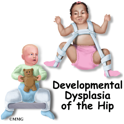
Developmental Dysplasia of the Hip


Developmental dysplasia of the hip (DDH) is a deformity of the hip that can occur before, during, or weeks after birth.

At periodic checkups, a doctor will examine your baby's hips to rule out DDH, which can cause hip dislocation and/or an abnormal walk. It's important to recognize DDH early, so a child can receive timely treatment and avoid orthopedic problems later in life.

What Is DDH?

The hip is a ball-and-socket joint. In a normal-functioning hip, the rounded top of the thighbone, or femoral head, rests comfortably in the acetabulum (the cup-like hipbone socket).

In mild cases of DDH, the femoral head moves back and forth within the socket, causing a child to have an unstable hip. In more serious cases, the head becomes dislocated, moving completely out of the socket, but sometimes can be put back in with pressure. In the most severe cases, the femoral head may not even reach the socket where it should be held in place.

Hip dislocations are relatively uncommon, affecting just 1 in 1,000 live births. However, some degree of instability of the hip is seen in as many as 1 in 3 newborns. Girls are more likely to develop dislocations of the hip.

What Causes It?

The causes of DDH aren't completely understood, but experts think that many factors are involved. The cramping of the fetus inside the uterus — which is more likely to happen in first pregnancies when the uterus is tight, or in pregnancies where there is a decrease of amniotic fluid (liquid in the womb) — can increase the likelihood of DDH.

Other factors include abnormal positioning of the fetus inside the womb, such as being in the breech position (buttocks face the birth canal), especially when the knees extend out with the feet near the head (called "frank breech"). Having other conditions develop as a result of positioning, like metatarsus adductus (an inward curving of the foot), increases the odds of a child developing DDH.

DDH also may be caused by the infant's response to the mother's hormones that relax the ligaments for labor and delivery, causing the baby's hip to soften and stretch during labor. In 20% of cases, family history is a factor, and if it is, future children should be checked by ultrasound at 6 weeks of age.

Signs and Symptoms

DDH usually affects only one side of the body, most often the right side, and pain is rare.

Infants often don't show signs that they have DDH, and there may be no signs at all. Still, doctors look for these indicators:

  • at birth, an inability to move the thigh outward at the hip as far as possible
  • an audible "click" during routine post-natal checkups
  • different leg lengths
  • asymmetry in the fat folds of the thigh around the groin or buttocks
  • after 3 months, asymmetry in the motion of the hip and obvious shortening of the affected leg
  • in older kids, an exaggeration in the spinal curvature that may develop to compensate for the abnormally developed hip
  • limping in older children

Diagnosis

A doctor can determine whether a hip is dislocated or likely to become dislocated by gently pushing and pulling on the child's thighbones, and determining whether they are loose in their sockets. In one commonly used diagnostic test, a child lies on a flat surface and his or her thighs are spread out in order to gauge the hips' range of motion.

A second test brings the knees together and attempts to push the femoral head rearward, out of the socket. It is during these tests that the doctor will hear a "click," which may indicate a dislocation. These maneuvers are done at routine checkups until the baby is walking normally.

Sometimes a doctor will recommend that a child undergo an X-ray or ultrasound to get a better view of a dislocated hip. Ultrasounds are recommended for babies under 4 months of age, while X-rays are performed on older kids. Prior to 4 months of age, hip tissue has not yet hardened (or ossified) from flexible cartilage to bone, and therefore does not show up on X-ray images that capture only bony anatomy and not cartilage.

Treatment

Treatment for DDH depends on the age of the child and the severity of the condition. Mild cases may correct themselves in the first few weeks of life.

If an unstable hip is seen in newborns, the hip may be held in position with a Pavlik harness. This device keeps the femoral head in its socket by holding the knee up toward the child's head. A shoulder harness attaches to foot stirrups to keep the leg elevated. The goal is to tighten the ligaments in the area and stimulate normal forming of the hip socket. Treatment with the Pavlik harness lasts about 6 to 12 weeks, and continues until the hip is stable and ultrasound exams are normal.

After a child reaches 6 months of age the Pavlik harness will be ineffective. Older kids will need to undergo one of the following treatments:

  • Closed reduction. The bone is manually put back into place after the child is put under anesthesia. This treatment is usually preferred in children younger than 18 months.
  • Open reduction. The hip is realigned and the thighbone is placed back into the hip socket through surgery. During the procedure, tight muscles and tissues surrounding the hip joint are loosened and then later tightened up once the hip is back in place. This is the procedure of choice for kids older than 18 months.

After reaching age 2 or 3, a child might need surgery on the pelvis to deepen the hip socket (if it's too shallow) or to shorten the thighbone or realign it. Following surgery, the child is put in a hip spica (body cast). About 1 in 20 babies with DDH needs more than the Pavlik harness to correct the condition.

Caring for Your Child

DDH can't be prevented, but if it's recognized early and treated appropriately, most children will develop normally and have no related problems. DDH does not cause pain initially, but if left untreated it can result in significant impairment of function.

Kids with untreated DDH will have legs of uneven lengths in adulthood, which can lead to a limp or waddling gait, back and hip pain, and overall decreased agility.

Hydrocephalus


Hydrocephalus is a brain condition that gets its name from the Greek word for water (meaning "hydro") and head (meaning "cephalus"). It occurs when cerebral spinal fluid (CSF) — the clear, water-like fluid that surrounds and cushions the brain and spinal cord — is unable to drain from the brain. It then pools, causing a backup of fluid in the skull.

Sometimes referred to as "water on the brain," hydrocephalus can cause babies' and young children's heads to swell to accommodate the excess fluid. Older kids, whose skull bones have matured and fused together, experience painful headaches due to increased pressure in the head.

If left untreated, hydrocephalus can lead to brain damage, a loss in mental and physical abilities, and even death. With early diagnosis and timely treatment, however, most children recover successfully.

Causes of Hydrocephalus

When everything is working normally inside the brain, CSF will flow through narrow passageways called ventricles and exit the brain through a small reservoir at the base of the brain called the cistern. CSF is responsible for delivering nutrients to the brain and taking waste away from sensitive areas, where it will eventually be absorbed into the bloodstream.

If a blockage exists in any of the ventricles, CSF backs up and causes an excess of fluid in the brain, or hydrocephalus. This accumulation of fluid can also happen when the choroid plexus (the area of the brain that produces CSF) is in overdrive or if the fluid fails to be properly absorbed by the bloodstream.

When hydrocephalus is present at birth, it can be the result of conditions like spina bifida (where the primary cause is abnormal development of the spinal cord) or aqueductal stenosis (a narrowing of the small passageway, called the "aqueduct of Sylvius," that connects two major ventricles in the brain).

Acquired hydrocephalus is caused by cranial hemorrhage, or bleeding in the brain. This can happen in premature babies or kids who've undergone traumatic head injuries.

But not every case of hydrocephalus is due to the causes mentioned here: Some kids can develop hydrocephalus without a known cause.

Signs in Babies

Symptoms of hydrocephalus vary depending on the age of a child.

Infants and babies under the age of 1 year will appear to have significant swelling of the head. Their skull bones — thin, bony plates that have not yet fused together — are connected by fibrous tissue called sutures. These sutures, or "soft spots," have not yet hardened and therefore stretch and protrude to accommodate the excess CSF.

As a result, a baby with hydrocephalus will appear to have an abnormally shaped head — usually much larger than other babies the same age. Other signs to look for include:

  • bulging at the soft spots
  • "split" sutures — a gap can be felt between skull bones
  • rapid increase in head circumference
  • swollen veins that are recognizable to the naked eye
  • downward cast of the eyes (called "sunsetting")

Depending on the severity of the condition, kids may also experience sleepiness, irritability, vomiting, and seizures. In extreme cases, a child may also experience "failure to thrive," meaning that he or she might miss growth or developmental milestones or may revert to earlier developmental stages.

Signs in Older Kids

Older children will not have the easily recognizable symptom of an enlarged head because their skull bones have fused together and therefore cannot expand to accommodate the excess fluid.

In these cases, pressure on the brain intensifies and causes severe headaches that may wake a child in the middle of the night or early in the morning. Headaches may be coupled with:

  • nausea/vomiting
  • sleepiness
  • difficulties with balance and motor skills
  • double vision
  • squinting and/or other repetitive eye movements
  • seizures

Changes in personality, loss of new developmental abilities (like speaking or walking), and memory loss may also occur in more advanced cases.

Diagnosis

A child who shows any of the signs and symptoms mentioned above should be evaluated by a doctor right away. The doctor will perform examinations, which may include a medical history and diagnostic imaging like ultrasound, CT (computed tomography), and MRI (magnetic resonance imaging) to get a clear picture of the inside of the brain.

During imaging, a child will lie still on a table for a few seconds while a machine passes over his or her body, emitting very low (and harmless) frequencies of radiation or sound waves.

Shunt Procedure

If hydrocephalus is diagnosed, treatment will depend on the age of the child, the cause of the cerebral spinal fluid build-up (whether from a blockage, overproduction of fluid, or another problem), and a child's overall health.

Shunt procedures, which have been the standard of care for decades, involve surgically implanting one end of a catheter (flexible tube) into a ventricle of the brain and placing the other end in the abdominal cavity, chambers of the heart, or space around the lungs where fluid is drained and absorbed by the bloodstream. A valve in the shunt system regulates flow to prevent over-draining and under-draining.

While shunting is often an effective treatment for hydrocephalus, there is a high chance of failure and complications. About 30% of shunts will stop working within the first year, with about 5% failing in each subsequent year, causing symptoms to recur. A child will need to have surgery to correct the problem — whether it requires replacing a catheter or valve or replacing the entire shunt. Most kids who undergo shunting will require subsequent operations over their lifetimes to regulate shunt problems.

Infections are another side effect of shunting, and occur in 5%-10% of shunt operations. Kids will develop typical signs of infection, like fever and neck stiffness, and may feel tenderness along the shunt or belly pain. Most infections develop within the first several months after a shunt procedure and require temporary removal of the device while a child receives intravenous antibiotics for up to 2 weeks.

Ventriculectomy

A second, and increasingly more common, treatment for hydrocephalus is an endoscopic third ventriculectomy. During this procedure, a small opening is made in the bottom of the third ventricle (one of four ventricles in the brain) to allow fluid to exit the brain.

This minimally invasive approach involves placing an endoscope (small lighted camera) inside the brain to provide surgeons with a view of the surgical site on a computer monitor. Then, using very small instruments, the doctor will make a tiny hole in the bottom of the third ventricle, where a thin membrane separates the inside and outside of the brain. This new "evacuation route" permits fluid to drain normally into the spaces outside the brain while bypassing any obstructions that are causing a backup, so the body can reabsorb the CSF back into the bloodstream as it normally would.

When deemed appropriate, third ventriculectomies are the procedure of choice for kids older than 6 months of age due to a higher efficacy rate and lower risk of infection than shunting. Those who undergo the procedure have up to a 90% chance of long-term success, with little need for follow-up procedures. Third ventriculectomies also have shown promise in newborns and may be offered as a treatment to these younger patients in the near future.

Outlook

With timely treatment, many kids with hydrocephalus go on to lead normal lives.

Those with more complex medical problems, like spina bifida or bleeding in the brain from prematurity, may experience a higher rate of complications due to their underlying medical conditions. In these kids, early intervention greatly improves the chances of recovery.