Pyloric Stenosis

While you were anticipating your new baby, you probably mentally prepared yourself for the messier aspects of child rearing: poopy diapers, food stains, and of course, spit up. But what's normal and what's not when it comes to spitting up or vomiting in infants?

Pyloric stenosis, a condition that affects the gastrointestinal tract during infancy, isn't normal - it can cause your baby to vomit forcefully and often and may cause other problems such as dehydration and salt and fluid imbalances. Keep reading to understand why getting immediate treatment for pyloric stenosis is so important.

What Is Pyloric Stenosis?

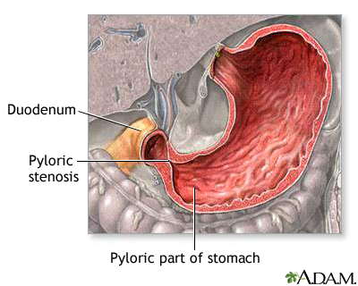
Pyloric stenosis is a narrowing of the pylorus, the lower part of the stomach through which food and other stomach contents pass to enter the small intestine. When an infant has pyloric stenosis, the muscles in the pylorus have become enlarged to the point where food is prevented from emptying out of the stomach.

Also called infantile hypertrophic pyloric stenosis or gastric outlet obstruction, pyloric stenosis is fairly common - it affects about three out of 1,000 babies in the United States. Pyloric stenosis is about four times more likely to occur in firstborn male infants. It has also been shown to run in families - if a parent had pyloric stenosis, then an infant has up to a 20% risk of developing the condition. Pyloric stenosis occurs more commonly in Caucasian infants than in babies of other ethnic backgrounds, and affected infants are more likely to have blood type B or O.

Most infants who develop pyloric stenosis are usually between 2 weeks and 2 months of age - symptoms usually appear during or after the third week of life. It is one of the more common causes of intestinal obstruction during infancy that requires surgery.

What Causes Pyloric Stenosis?

It is believed that babies who develop the condition are not born with pyloric stenosis, but that the progressive thickening of the pylorus occurs after birth. An affected infant begins showing symptoms when the pylorus is so thickened that the stomach can no longer empty properly.

It is not known exactly what causes the thickening of the muscles of the pylorus - it may be a combination of several factors. Some researchers believe that maternal hormones could be a contributing cause. Others believe that the thickening of the muscle is the stomach's response to some type of allergic reaction in the body.

Some scientists believe that babies with pyloric stenosis lack receptors in the pyloric muscle that detect nitric oxide, a chemical in the body that tells the pylorus muscle to relax. As a result, the muscle is in a state of contraction almost continually, which causes it to become larger and thicker over time. It may take some time for this thickening to occur, which is why pyloric stenosis usually appears in babies a few weeks after birth.

Signs and Symptoms

Symptoms of pyloric stenosis generally begin around 3 weeks of age. They include:

  • Vomiting - The first symptom of pyloric stenosis is usually vomiting. At first it may seem that the baby is simply spitting up frequently, but then it tends to progress to projectile vomiting, in which the breast milk or formula is ejected forcefully from the mouth, in an arc, sometimes over a distance of several feet. Projectile vomiting usually takes place soon after the end of a feeding, although in some cases it may be delayed for hours. Rarely, the vomit may contain blood.

    In some cases, the vomited milk may smell curdled because it has mixed with stomach acid. The vomit will not contain bile, a greenish fluid from the liver that mixes with digested food after it leaves the stomach.

    Despite vomiting, a baby with pyloric stenosis is usually hungry again soon after vomiting and will want to eat. The symptoms of pyloric stenosis can be deceptive because even though a baby may seem uncomfortable, he may not appear to be in great pain or at first look very ill.
  • Changes in stools - Babies with pyloric stenosis usually have fewer, smaller stools because little or no food is reaching the intestines. Constipation or stools that have mucus in them may also be symptoms.
  • Failure to gain weight and lethargy - Most babies with pyloric stenosis will fail to gain weight or will lose weight. As the condition worsens, they are at risk for developing fluid and salt abnormalities and becoming dehydrated.

    Dehydrated infants are lethargic and less active than usual, and they will develop a sunken "soft spot" on their heads, sunken eyes, and a doughy, softened, or wrinkled appearance of the skin on the belly and upper parts of the arms and legs. Because urine output is decreased, it may be more than 4 to 6 hours between wet diapers.

    After feeds, increased stomach contractions may make noticeable ripples, or waves of peristalsis, which move from left to right over the infant's belly as the stomach tries to empty itself against the thickened pylorus.

It's important to talk to your child's doctor if your baby experiences any of these symptoms.

Other conditions can have similar symptoms as pyloric stenosis. For instance, gastroesophageal reflux disease (GERD) usually begins before 8 weeks of age, with excess spitting up, or reflux - which may resemble vomiting - taking place after feedings. However, most infants with GERD do not experience projectile vomiting, and although they may have poor weight gain, they tend to have normal stools.

In infants, symptoms of gastroenteritis - inflammation in the digestive tract that can be caused by viral or bacterial infection - may also somewhat resemble pyloric stenosis. Vomiting and dehydration are seen with both conditions; however, infants with gastroenteritis usually also have diarrhea with loose, watery, or sometimes bloody stools. Diarrhea usually isn't seen with pyloric stenosis.

Diagnosis and Treatment

Your child's doctor will ask detailed questions about the baby's feeding and vomiting patterns, including the appearance of the vomit. The most important part of diagnosing pyloric stenosis is a reliable and consistent history and description of the vomiting.

The baby will be fully examined, and any weight loss or failure to maintain growth since birth will be noted. During the exam, the doctor will attempt to feel if there is a pyloric mass - a firm, movable lump that feels like an olive and is sometimes detected in the belly of an infant with pyloric stenosis. If the doctor feels this mass, it's a strong indication that the baby has pyloric stenosis; the baby will be referred to a pediatric surgeon and hospitalized for further treatment.

If the baby's feeding history and physical examination suggest pyloric stenosis but no "olive" is felt, then an ultrasound of the baby's abdomen will usually be performed. The enlarged, thickened pylorus can be seen on ultrasound images.

Sometimes instead of an ultrasound, a barium swallow is performed. The baby swallows a small amount of a chalky liquid (barium), and then special X-rays are taken to view the pyloric region of the stomach to see if there is any narrowing or obstruction.

Infants suspected of having pyloric stenosis usually undergo blood tests because the continuous vomiting of stomach acid, as well as the resulting dehydration from fluid losses, can cause salt (electrolyte) imbalances in the blood that need to be corrected.

When an infant is diagnosed with pyloric stenosis, either through physical examination, ultrasound, or barium swallow, the baby will be admitted to the hospital and prepared for surgery. Any dehydration or electrolyte problems in the blood will be corrected with intravenous (IV) fluids, usually within 24 hours.

A surgical procedure called pyloromyotomy, which involves cutting through the thickened muscles of the pylorus, is performed to relieve the obstruction from pyloric stenosis. The pylorus is examined through a very small incision, and the muscles that are overgrown and thickened are spread. Nothing is cut out - the stitches are under the skin and there are no stitches or clips to remove.

After surgery, most babies are able to return to normal feedings fairly quickly. The baby starts feeding again 3 to 4 hours after the surgery, and the baby can return to breast-feeding or the formula that he was on prior to the surgery. Because of swelling at the surgery site, the baby may still vomit small amounts for a day or so after surgery. As long as there are no complications, most babies who have undergone pyloromyotomy can return to a normal feeding schedule and be sent home within 48 hours of the surgery.

If you are breast-feeding, you may be concerned about being able to continue feeding while your baby is hospitalized. The hospital should be able to provide you with a breast pump and assist you in its use so that you can continue to express milk until your baby can once again feed regularly.

After a successful pyloromyotomy, your infant will not need to follow any special feeding schedules. Your child's doctor will probably want to examine your child at a follow-up appointment to make sure the surgical site is healing properly and that your infant is feeding well and maintaining or gaining weight.

Pyloric stenosis should not recur after a complete pyloromyotomy. If your baby continues to display symptoms weeks after the surgery, it may suggest another medical problem, such as inflammation of the stomach (gastritis) or GERD - or it could indicate that the initial pyloromyotomy was incomplete.

When to Call Your Child's Doctor

Pyloric stenosis is a medical emergency that requires immediate treatment. Call your child's doctor if your baby has any of the following symptoms:

  • persistent or projectile vomiting after feeding
  • poor weight gain or weight loss
  • decreased activity or lethargy
  • few or no stools over a period of 1 or 2 days
  • signs of dehydration such as decreased urination (more than 4 to 6 hours between wet diapers); wrinkly or doughy appearance of the skin on the arms, legs, or belly; sunken "soft spot" on the head; sunken eyes; or jaundice (yellowing of the skin)

Osteosarcoma

Osteosarcoma is the most common type of bone cancer, and the sixth most common type of cancer in children. Although other types of cancer can eventually spread to parts of the skeleton, osteosarcoma is one of the few that actually begin in bones and sometimes spread (or metastasize) elsewhere, usually to the lungs or other bones.

Because osteosarcoma usually develops from osteoblasts (the cells that make growing bone), it most commonly affects teens who are experiencing a growth spurt. Boys are more likely to have osteosarcoma than girls, and most cases of osteosarcoma involve the knee.

Most osteosarcomas arise from random and unpredictable errors in the DNA of growing bone cells during times of intense bone growth. There currently isn't an effective way to prevent this type of cancer. But with the proper diagnosis and treatment, most kids with osteosarcoma do recover.

Risk for Childhood Osteosarcoma

Osteosarcoma is most often seen in teenage boys, and evidence shows that teens who are taller than average have an added risk for developing the disease.

Kids who have inherited one of the rare cancer syndromes also are at higher risk for osteosarcoma. These syndromes include retinoblastoma (a malignant tumor that develops in the retina, usually in children younger than age 2) and Li-Fraumeni syndrome (a kind of inherited genetic mutation). Because exposure to radiation is another trigger for DNA mutations, children who have received radiation treatments for a prior episode of cancer are also at increased risk for osteosarcoma.

Symptoms of Osteosarcoma

The most common symptoms of osteosarcoma are pain and swelling in a child's leg or arm. It occurs most often in the longer bones of the body — such as above or below the knee or in the upper arm near the shoulder. Pain may be worse during exercise or at night, and a lump or swelling may develop in the affected area up to several weeks after the pain starts. Pain that persistently wakes the child up at night or pain at rest are of particular concern. In osteosarcoma of the leg, the child may also develop an unexplained limp. In some cases, the first sign of the disease is a broken arm or leg, because the cancer has weakened the bone to make it vulnerable to a break.

If your child or teen has any of the above symptoms, it's important to see a doctor.

Diagnosing Osteosarcoma

To diagnose osteosarcoma, the doctor will likely perform a physical exam, obtain a detailed medical history, and order X-rays to detect any changes in bone structure. The doctor may also order a magnetic resolution imaging (MRI) scan of the affected area, which will find the best area to biopsy and show whether the osteosarcoma has spread from the bone into nearby muscles and fat. The doctor will also order a bone biopsy to obtain a sample of the tumor for examination in the lab. This is best done by an orthopedic surgeon experienced in the treatment of osteosarcoma (orthopedic oncologist).

Sometimes the doctor does a needle biopsy, using a long hollow needle to take a sample of the tumor. A local anesthesia is typically used in the area that's being biopsied. Alternatively, the doctor may order an open biopsy, in which a portion of the tumor is removed in the operating room by a surgeon while the child is under general anesthesia.

If a diagnosis of osteosarcoma is made, the doctor will order CT chest scans as well as a bone scan and, sometimes, additional MRI studies. These will show if the cancer has spread to any part of the body beyond the original tumor. These tests will be repeated after treatment starts to determine how well it is working and whether the cancer is continuing to spread.

Treating Osteosarcoma

Treatment of osteosarcoma in children includes chemotherapy (the use of medical drugs to kill cancer cells and shrink the cancer) followed by surgery (to remove cancerous cells or tumors) and then more chemotherapy (to kill any remaining cancer cells and minimize chances of the cancer coming back). Surgery often can effectively remove bone cancer, while chemotherapy can help eliminate remaining cancer cells in the body.

Surgical Treatment

Surgical treatments for osteosarcoma consist of either amputation or limb-salvage surgery.

Currently, most teens with osteosarcomas involving an arm or leg can be treated with limb-salvage surgery rather than amputation. In limb-salvage surgery, the bone and muscle affected by the osteosarcoma are removed, leaving a gap in the bone that is filled by either a bone graft (usually from the bone bank) or more often a special metal prosthesis. These can be appropriately matched to the size of the bone defect. The risk of infection and fracture is higher with bank bone replacement and therefore metal prostheses are more commonly used for reconstruction of the bone after removal of the tumor.

If the cancer has spread to the nerves and blood vessels surrounding the original tumor on the bone, amputation (removing part of a limb along with the osteosarcoma) is often the only choice.

When osteosarcoma has spread to the lungs or elsewhere, surgery may also be performed to remove tumors in these distant locations.

Chemotherapy

Chemotherapy is usually given both before and after surgery. It eliminates small pockets of cancer cells in the body, even those too small to appear on medical scans. A child or teen with osteosarcoma is given the chemotherapy drugs intravenously (through a vein) or orally (by mouth). The drugs enter the bloodstream and work to kill cancer in parts of the body where the disease has spread, such as the lungs or other organs.

Short-Term and Long-Term Side Effects

Amputation carries its own short-term and long-term side effects. It usually takes at least 3 to 6 months until a young person learns to use a prosthetic (artificial) leg or arm, and this is just the beginning of long-term psychological and social rehabilitation.

With a limb salvage surgery, one usually starts bending the knee or the affected body part almost immediately. A continuous passive motion (CPM) machine, that continuously bends and straightens the knee may be used to improve motion for tumors around the knee. Physical therapy and rehabilitation for 6 to 12 months following surgery usually enables the child to walk initially with a walker or crutches and then without any assistive devices. Early complications after surgery include infection and slow healing of the surgical wound, and the metal prosthetic device or the bank bone may need to be replaced in the long term. Other late problems might include fracture of the bank bone or failure of the bank bone to heal to the child's bone, which might require more surgery.

Many of the medications used in chemotherapy also carry the risk of both short-term and long-term problems. Short-term effects include anemia, abnormal bleeding, and increased risk of infection due to destruction of the bone marrow, as well as kidney damage and menstrual irregularities. Some drugs carry a risk of bladder inflammation and bleeding into the urine, hearing loss, and liver damage. Others may cause heart and skin problems. Years after chemotherapy for osteosarcoma, patients have an increased risk of developing other cancers.

Chances for a Cure

Recent studies have reported that survival rates of 60% to 80% are possible for osteosarcoma that hasn't spread beyond the tumor, depending on the success of chemotherapy.

Osteosarcoma that has spread cannot always be treated as successfully. Also, a child whose osteosarcoma is located in an arm or leg generally has a better prognosis than one whose disease involves the ribs, shoulder blades, spine, or pelvic bones.

New Treatments

Treatments are being developed and researched with new chemotherapy drugs. Other research is focused on the role certain growth factors might play in the development of osteosarcoma. This research may be used to develop new medications to slow these growth factors as a way to treat the cancer.

For osteosarcomas that cannot be removed surgically, studies are now underway to test treatments that use new combinations of chemotherapy and localized, high-dose radiation.

Peptic Ulcers

Many people think that spicy foods cause ulcers, but the truth is that bacteria called Helicobacter pylori (or H. pylori) are the main culprit. And while many believe that adults in high-stress jobs are the only ones affected, people of any age — even children — can develop ulcers. Doctors say that in the United States, almost 1 in every 10 people will get an ulcer at some time during their lives.

What Are Peptic Ulcers?

An ulcer is a sore, which means it's an open, painful wound. Peptic ulcers are ulcers that form in the stomach or the upper part of the small intestine, called the duodenum. An ulcer in the stomach is called a gastric ulcer and an ulcer in the duodenum is called a duodenal ulcer.

Both a gastric ulcer and a duodenal ulcer result when H. pylori or a drug weakens the protective mucous coating of the stomach and duodenum, allowing acid to get through to the sensitive lining beneath. Both the acid and the bacteria can irritate the lining and cause an ulcer to form.

H. pylori infection is usually contracted in childhood, perhaps through food, water, or close contact with an infected individual. Infections are more common in adults older than age 60 and in developing countries. And most people with H. pylori don't display any symptoms until they're older. In fact, they may go through life unaware that they're infected.

Although H. pylori infection usually doesn't cause problems in childhood, if left untreated it can cause gastritis (the irritation and inflammation of the lining of the stomach), peptic ulcer disease, and even stomach cancer later in life.

In the past, having peptic ulcers meant living with a chronic condition for several years or even a lifetime. But today, a better understanding of the cause of peptic ulcers and how to treat them means that most people can be cured.

What Causes Peptic Ulcers in Children?

Although stress and certain foods may aggravate an ulcer, most ulcers are caused by an H. pylori infection or the use of common nonsteroidal anti-inflammatory drugs (NSAIDs) such as ibuprofen.

However, whereas most experts agree that H. pylori infection is a primary cause of peptic ulcers in adults, not everyone thinks that the bacteria are a major culprit in childhood ulcers. Some doctors make the distinction between duodenal ulcers, which are commonly associated with H. pylori infection, and gastric ulcers, which may stem from other causes.

It's recognized that certain medical conditions can contribute to the development of ulcers. For instance, children with severe burns can develop ulcers secondary to the stress of their injuries. This is also true for infants who become septic, or very ill with a bacterial infection. In otherwise healthy children, peptic ulcers are very unusual.

Some doctors believe that more children get drug-related gastric ulcers than other types of peptic ulcers. Even moderate use of NSAIDs can cause gastrointestinal problems and bleeding in some children. Acetaminophen does not cause stomach ulcers and is a good alternative to NSAIDs for kids.

What Are the Signs and Symptoms?

Although peptic ulcers are rare in children, if your child exhibits any of the following signs and symptoms, you should call your child's doctor:

  • burning pain in the abdomen between the breastbone and the belly button (the most common ulcer symptom)
  • nausea
  • vomiting
  • chest pain (usually dull and achy)
  • loss of appetite
  • frequent burping or hiccuping
  • weight loss
  • feeding difficulties
  • blood in vomit or bowel movements, which may appear dark red or black

These signs and symptoms are common in many childhood illnesses and don't necessarily mean that your child has an ulcer, but they should be reported to your child's doctor. Based on your child's medical history and symptoms, the doctor may refer your child to a pediatric gastroenterologist (a doctor who specializes in disorders of the stomach, intestines, and associated organs) for further evaluation.

How Are Peptic Ulcers Diagnosed?

The doctor may do an upper gastrointestinal (GI) series to get a close look at your child's gastrointestinal tract. An upper GI series is a set of X-rays of the esophagus, stomach, and duodenum.

The doctor may also order an upper endoscopy, especially if an ulcer is suspected. This procedure is performed under sedation and involves inserting an endoscope — a small, flexible tube with a tiny camera on the end — down your child's throat and into the stomach and duodenum. It allows the doctor to see the lining of the esophagus, stomach, and duodenum to check for possible ulcers, inflammation, or food allergies. The endoscope can also be used to perform tissue tests to detect the presence of H. pylori.

The endoscopy is often used in conjunction with a test called a pH probe in which a small wire is inserted into the lower part of the esophagus to measure the amount of acid going into that area.

If an ulcer is found, your child's doctor will test for H. pylori. This test is important because treatment for an ulcer caused by H. pylori is different from the treatment for an ulcer caused by NSAIDs.

H. pylori may be diagnosed through:

  • tissue tests (performed during an endoscopy)
  • blood tests (which can detect the presence of H. pylori antibodies; blood tests are common, although they typically aren't as accurate for children as they are for adults)
  • stool tests (which can detect the presence of H. pylori antigens; stool tests are becoming more common for detecting H. pylori, and some doctors think they're more accurate than blood tests)
  • breath tests (which can detect carbon broken down by H. pylori after the patient drinks a solution; breath tests are also used mostly in adults)

How Are Peptic Ulcers Treated?

The good news is that most H. pylori-related ulcers are curable with treatment that combines two different kinds of antibiotics and an acid suppressor. The medication is taken over a 1- to 2-week period. The ulcer may take 8 weeks to heal, but the pain usually goes away after a few days or a week. To be sure the treatment has worked, doctors may do a follow-up endoscopy 6 to 12 months later to check for H. pylori.

Likewise, ulcers related to NSAIDs rarely require surgery and usually improve with an acid suppressor and stopping or changing the NSAID. No antibiotics are needed to treat this type of ulcer.

Caring for Your Child

If your child is diagnosed with an H. pylori-related ulcer, make sure he or she takes all of the antibiotics as directed by the doctor. Even if the symptoms disappear, the infection may not be gone until all of the medication has been taken.

If your child has a medication-related ulcer, the doctor will tell you to avoid NSAIDs, including any medication containing ibuprofen or aspirin. Also, be sure to give your child the prescribed acid-reducing medication.

Unless a particular food is bothersome, most doctors don't recommend dietary restrictions for children with ulcers. A good diet with a variety of foods is essential to a child's growth and development.

Alcohol and smoking can aggravate an ulcer. Also make sure that your child avoids coffee, tea, sodas, and foods that contain caffeine, which can stimulate the secretion of acid in the stomach and may make an ulcer worse.

When Should You Call Your Child's Doctor?

Call your child's doctor immediately if your child displays any of these symptoms:

  • sudden, sharp, persistent belly pain
  • bloody or black bowel movements
  • bloody vomit or vomit that looks like coffee grounds

If your child has peptic ulcer disease, these signs and symptoms could indicate a serious problem, such as:

  • perforation (when the ulcer becomes too deep and breaks through the stomach or duodenal wall)
  • bleeding (when acid or the ulcer breaks a blood vessel)
  • obstruction (when the ulcer blocks the path of food from going through the intestines)

If your child is taking NSAIDs and experiences symptoms of peptic ulcer disease, seek prompt medical attention. Delaying diagnosis and treatment can lead to complications and possibly the need for surgery. But with timely treatment, almost all peptic ulcers can be cured.

Necrotizing Enterocolitis

As if the birth of a premature baby isn't stressful enough, there are a number of diseases premature babies can experience within the first weeks of life. Necrotizing enterocolitis (NEC) is one of them. "Necrotizing" means the death of tissue, "entero" refers to the small intestine, "colo" to the large intestine, and "itis" means inflammation. But knowing what the words mean is only the start of understanding this infant disease.

What Is Necrotizing Enterocolitis?

A gastrointestinal disease that mostly affects premature infants, NEC involves infection and inflammation that causes destruction of the bowel (intestine) or part of the bowel. Although it affects only one in 2,000 to 4,000 births, or between 1% and 5% of neonatal intensive care unit (NICU) admissions, NEC is the most common and serious gastrointestinal disorder among hospitalized preterm infants.

NEC typically occurs within the first 2 weeks of life, usually after milk feeding has begun (at first, feedings are usually given through a tube that goes directly to the baby's stomach). About 10% of babies weighing less than 1,500 grams (3 lbs., 5 oz.) experience NEC. These premature infants have immature bowels, which are sensitive to changes in blood flow and prone to infection. They may have difficulty with blood and oxygen circulation and digestion, which increases their chances of developing NEC.

What Causes It?

The exact cause of NEC is unknown, but several theories exist. It is thought that the intestinal tissues of premature infants are weakened by too little oxygen or blood flow, and when feedings are started, the added stress of food moving through the intestine allows bacteria that are normally found in the intestine to invade and damage the wall of the intestinal tissues. The damage may affect only a short segment of the intestine, or it may progress quickly to involve a much larger portion.

The infant is unable to continue feedings and starts to appear ill if bacteria continues to spread through the wall of the intestines and sometimes into the bloodstream. He may also develop imbalances in the minerals in the blood. In severe cases of NEC, a hole (perforation) may develop in the intestine, allowing bacteria to leak into the abdomen and causing life-threatening infection (peritonitis). Because the infant's body systems are immature, even with quick treatment for NEC there may be serious complications.

Other factors seem to increase the risk of developing NEC. Some experts believe that the makeup of infant formula, the rate of delivery of the formula, or the immaturity of the mucous membranes in the intestines can cause NEC. (Babies who are fed breast milk can also develop NEC, but their risk is lower.) Another theory is that babies who have had difficult deliveries with lowered oxygen levels can develop NEC. When there is not enough oxygen, the body sends the available oxygen and blood to vital organs instead of the gastrointestinal tract, and NEC can result. Babies with an increased number of red blood cells (polycythemia) in the circulation also seem to be at higher risk for NEC. Too many red blood cells thicken the blood and hinder the transport of oxygen to the intestines.

NEC sometimes seems to occur in "epidemics," affecting several infants in the same nursery. Although this may be due to coincidence, it suggests the possibility that it could in some cases be spread from one baby to another, despite the fact that all nurseries have very strict precautions to prevent the spread of infection.

Signs and Symptoms

The symptoms of NEC may resemble other digestive conditions. Every infant experiences the symptoms of NEC differently, which may include:

  • poor tolerance to feedings
  • feedings stay in stomach longer than expected
  • decreased bowel sounds
  • abdominal distension (bloating) and tenderness
  • greenish (bile-colored) vomit
  • redness of the abdomen
  • increase in stools, or lack of stools
  • bloody stools

More subtle signs of NEC might include apnea (periodic stoppage of breathing), bradycardia (slowed heart rate), diarrhea, lethargy, and fluctuating body temperature. Advanced cases may show fluid in the peritoneal (abdominal) cavity, peritonitis (infection of the membrane lining the abdomen), or shock.

Diagnosis and Treatment

The diagnosis of NEC is usually confirmed by the presence of an abnormal gas pattern as seen on an X-ray. This is indicated by a "bubbly" appearance of gas in the walls of the intestine, large veins of the liver, or the presence of air outside of the intestines in the abdominal cavity. A surgeon may insert a needle into the abdominal cavity to withdraw fluid to determine whether there is a hole in the intestines.

The majority of infants with NEC are treated medically, and symptoms resolve without the need for surgery. Treatment includes:

  • stopping feedings
  • nasogastric drainage (inserting a tube through the nasal passages down to the stomach to remove air and fluid from the stomach and intestine)
  • intravenous fluids for fluid replacement and nutrition
  • antibiotics for infection
  • frequent examinations and X-rays of the abdomen

The baby's belly size is measured and watched carefully, and periodic blood samples are taken to determine the presence of bacteria. Stools are also checked for blood. If the abdomen is so swollen that it interferes with breathing, extra oxygen or mechanically assisted breathing (a ventilator) is used to help the baby breathe.

If the infant responds favorably, he may be back on regular feedings within 72 hours, although in most cases feedings are withheld and antibiotics are continued for 7 to 10 days. If the bowel perforates (tears) or the condition worsens, surgery may be indicated. Severe cases of NEC may require removal of a segment of intestine. Sometimes after removal of diseased bowel, the healthy areas can be sewn back together. Other times, especially if the baby is very ill or there is spillage of stool in the abdomen, the surgeon will bring an area of the intestine or bowel to an opening on the abdomen (called an ostomy).

Most infants who develop NEC recover fully and do not have further feeding problems. In some cases, scarring and narrowing of the bowel may occur and can cause future intestinal obstruction or blockage. Another residual problem may be malabsorption (the inability of the bowel to absorb nutrients normally). This is more common in children who required surgery for NEC and had part of their intestine removed.

Caring for Your Child

NEC can be extremely frightening to parents. Parents who are deprived of the experience of feeding their babies will certainly feel frustrated - their infant is so small, it just doesn't feel right to stop feeding him. As important as it is to be able to hold and bond with your baby, this may not be possible while the baby is in critical condition.

Listen to and take comfort from the NICU staff - they are trained and eager to support parents of preemies as well as the preemies themselves. Remember that there's a good chance that your baby will be back on regular feedings within a short time. By that time, you'll be more than ready to hold and caress him.

Intestinal Malrotation

An intestinal obstruction is a blockage of the digestive tract that prevents the proper passage of food. Some intestinal obstructions are present at birth, while others are caused by such problems as hernias, abnormal scar tissue growth after an abdominal operation, and inflammatory bowel disease (IBD).

Malrotation is twisting of the intestines (or bowel) caused by abnormal development while a fetus is in utero, and can cause obstruction. Malrotation occurs in 1 out of every 500 births in the United States.

Some children with intestinal malrotation are born with other associated conditions, including:

  • other defects of the digestive system
  • heart defects
  • abnormalities of other organs, including the spleen or liver

Some kids with malrotation never experience complications and are never diagnosed. But most develop symptoms during infancy, and the majority are diagnosed by 1 year of age. Although surgery is required to repair malrotation, most kids will go on to grow and develop normally after treatment.

What Is Malrotation?

The small and large intestines are the longest part of the digestive system. If stretched out to their full length, they would measure more than 20 feet long by adulthood, but because they're folded up, they fit into the relatively small space inside the abdomen. Malrotation occurs when the intestines don't position themselves normally during fetal development and aren't attached inside properly as a result. The exact reason this occurs is unknown.

When a fetus develops in the womb, the intestines start out as a small, straight tube between the stomach and the rectum. As this tube develops into separate organs, the intestines move into the umbilical cord, which supplies nutrients to the developing embryo.

Around the 10th week of pregnancy, the intestines move from the umbilical cord into the abdomen. When they don't properly turn after moving into the abdomen, malrotation occurs.

Malrotation in itself may not cause any problems. However, it can lead to other complications:

  • Bands of tissue called Ladd's bands may form, obstructing the first part of the small intestine (the duodenum).
  • In a condition called volvulus, the bowel twists on itself, cutting off the blood flow to the tissue and causing the tissue to die. The symptoms associated with volvulus, including pain and cramping, are often what lead to the diagnosis of malrotation.
  • Obstruction caused by volvulus or Ladd's bands is a potentially life-threatening problem. The bowel can stop functioning and intestinal tissue can die from lack of blood supply if an obstruction isn't recognized and treated. Volvulus, especially, is an emergency situation, with the entire small intestine in jeopardy.

Signs and Symptoms

One of the earliest signs of malrotation and volvulus is abdominal pain and cramping caused by the inability of the bowel to push food past the obstruction. When infants experience this cramping they may:

  • pull up their legs and cry
  • stop crying suddenly
  • behave normally for 15 to 30 minutes
  • repeat this behavior when the next cramp happens

Infants also may be irritable, lethargic, or have irregular stools.

Vomiting is another symptom of malrotation, and it can help the doctor determine where the obstruction is located. Vomiting that happens soon after the baby starts to cry often means the obstruction is in the small intestine; delayed vomiting usually means the blockage is in the large intestine. The vomit may contain bile (which is yellow or green in color) or may resemble feces.

Additional symptoms of malrotation and volvulus may include:

  • a swollen abdomen that's tender to the touch
  • diarrhea and/or bloody stools (or sometimes no stools at all)
  • irritability or crying in pain, with nothing seeming to help
  • rapid heart rate and breathing
  • little or no urine because of fluid loss
  • fever

Diagnosis

If volvulus or another intestinal blockage is suspected, the doctor will examine your child and then may order X-rays, a computed tomography (CT) scan, or an ultrasound of the abdominal area.

The doctor may use barium or another liquid contrast agent to see the X-ray or scan more clearly. The contrast can show if the bowel has a malformation and can usually determine where a blockage is located. Adults and older children usually drink barium in a liquid form. Infants may need to be given barium through a tube inserted from the nose into the stomach, or sometimes are given a barium enema, in which the liquid barium is inserted through the rectum.

Treatment

Treating significant malrotation almost always requires surgery. The timing and urgency will depend on the child's condition. If there is already a volvulus, surgery must be performed right away in order to prevent damage to the bowel.

Any child with bowel obstruction will need to be hospitalized. A tube called a nasogastric (NG) tube is usually inserted through the nose and down into the stomach to remove the contents of the stomach and upper intestines. This keeps fluid and gas from building up in the abdomen. Your child may also be given intravenous (IV) fluids to help prevent dehydration and antibiotics to prevent infection.

During the surgery, which is called a Ladd procedure, the intestine is straightened out, the Ladd's bands are divided, the small intestine is folded into the right side of the abdomen, and the colon is placed on the left side. Because the appendix is usually found on the left side of the abdomen when there is malrotation (it is normally found on the right), it is removed. Otherwise, should the child ever develop appendicitis, it could complicate diagnosis and treatment.

If it appears that blood may still not be flowing properly to the intestines, the doctor may perform a second surgery within 48 hours of the first. If the bowel still looks unhealthy at this time, the damaged portion may be removed.

If the child is seriously ill at the time of surgery, an ileostomy or colostomy will usually be performed. In this procedure, the diseased bowel is completely removed, and the end of the normal, healthy intestine is brought out through an opening on the skin of the abdomen (called a stoma). Fecal matter passes through this opening and into a bag that is taped or attached with adhesive to the child's belly. In young children, depending on how much bowel was removed, the ileostomy or colostomy is often a temporary condition that can later be reversed with another operation.

The majority of these surgeries are successful, although some children have recurring problems after surgery. Recurrent volvulus is rare, but a second bowel obstruction due to adhesions (scar tissue build-up after any type of abdominal surgery) could occur later.

Children who require removal of a large portion of the small intestine can have too little bowel to maintain adequate nutrition (a condition known as short bowel syndrome). They may be dependent on intravenous nutrition for a time after surgery (or even permanently if too little intestine remains) and may require a special diet afterward.

Most kids in whom the volvulus and malrotation are identified early, before permanent injury to the bowel has occurred, do well and develop normally.

If you suspect any kind of intestinal obstruction because your child has bilious (yellow or green) vomiting, a swollen abdomen, or bloody stools, call your doctor immediately, and take your child to the emergency room right away.

Inflammatory Bowel Disease

The digestive system is a set of organs (including the stomach, large and small intestines, rectum, and others) that convert the foods we eat into nutrients and absorb these nutrients into the bloodstream to fuel our bodies. We seldom notice its workings unless something goes wrong, as in the case of inflammatory bowel disease (IBD).

It's estimated that up to 1 million Americans have inflammatory bowel disease. It occurs most frequently in people ages 15 to 30, but it can also affect younger children and older people. And there are significantly more reported cases in western Europe and North America than in other parts of the world.

What Is Inflammatory Bowel Disease?

Inflammatory bowel disease (which is not the same thing as irritable bowel syndrome, or IBS) refers to two chronic diseases that cause inflammation of the intestines: ulcerative colitis and Crohn's disease. Although the diseases have some features in common, there are some important differences.

Ulcerative colitis is an inflammatory disease of the large intestine, also called the colon. In ulcerative colitis, the inner lining - or mucosa - of the intestine becomes inflamed (meaning the lining of the intestinal wall reddens and swells) and develops ulcers (an ulcer is a sore, which means it's an open, painful wound). Ulcerative colitis is often the most severe in the rectal area, which can cause frequent diarrhea. Mucus and blood often appear in the stool (feces or poop) if the lining of the colon is damaged.

Crohn's disease differs from ulcerative colitis in the areas of the bowel it involves - it most commonly affects the last part of the small intestine (called the terminal ileum) and parts of the large intestine. However, Crohn's disease isn't limited to these areas and can attack any part of the digestive tract. Crohn's disease causes inflammation that extends much deeper into the layers of the intestinal wall than ulcerative colitis does. Crohn's disease generally tends to involve the entire bowel wall, whereas ulcerative colitis affects only the lining of the bowel.

What Causes It?

Medical research hasn't determined yet what causes inflammatory bowel disease. But researchers believe that a number of factors may be involved, such as the environment, diet, and possibly genetics.

Current evidence suggests that there's likely a genetic defect that affects how our immune system works and how the inflammation is turned on and off in those people with inflammatory bowel disease, in response to an offending agent, like bacteria, a virus, or a protein in food.

The problem in people with the disease is that the inflammation gets turned on, but it doesn't get turned off. Medical evidence also indicates that smoking may enhance the likelihood of developing Crohn's disease.

What Are the Signs and Symptoms?

The most common symptoms of both ulcerative colitis and Crohn's disease are diarrhea and abdominal pain. Diarrhea can range from mild to severe (as many as 20 or more trips to the bathroom a day). If the diarrhea is extreme, it can lead to dehydration, rapid heartbeat, and a drop in blood pressure. And continued loss of small amounts of blood in the stool can lead to anemia.

At times, those with inflammatory bowel disease may also have constipation. With Crohn's disease, this can happen as a result of a partial obstruction (called stricture) in the intestines. In ulcerative colitis, constipation may be a symptom of inflammation of the rectum (also known as proctitis).

Because of the loss of fluid and nutrients from diarrhea and chronic inflammation of the bowel, someone with inflammatory bowel disease may also experience fever, fatigue, weight loss, dehydration, and malnutrition. Pain usually results from the abdominal cramping, which is caused by irritation of the nerves and muscles that control intestinal contractions.

But inflammatory bowel disease can cause other health problems that occur outside the digestive system. Although medical researchers don't know why these complications happen, some people with the disease may show signs of inflammation elsewhere in the body, such as in the joints, eyes, skin, and liver. Skin tags that look like hemorrhoids or abscesses may also develop around the anus.

Inflammatory bowel disease may also cause a delay in puberty or growth problems for some kids and teens with the condition, because it can interfere with a person getting nutrients from the foods he or she eats.

How Is It Diagnosed?

Inflammatory bowel disease can be hard to diagnose because there may be no symptoms, even if the person's bowel has become increasingly damaged for years. Once symptoms do appear, they often resemble those of other conditions, which may make it difficult for doctors to diagnose.

If your child has any of the symptoms of the disease, it's important to see your child's doctor. In addition to doing a physical examination, the doctor will ask you and your child about any concerns and symptoms your child has, your child's past health, your family's health, any medications your child is taking, any allergies your child may have, and other issues. This is called the medical history.

After hearing your child's symptoms, if the doctor suspects inflammatory bowel disease, he or she may suggest certain tests. Blood tests may be done to determine if there are signs of inflammation in your child's body, which are often present with the disease. The doctor may also check for anemia and for other causes of your symptoms, like infection.

The doctor will examine your child's stool for the presence of blood. He or she may look at your child's colon with an instrument called an endoscope. Also called a colonoscope or coloscope, this instrument is a long, thin tube inserted through the anus and attached to a TV monitor. This procedure is called a colonoscopy, which allows the doctor to see inflammation, bleeding, or ulcers on the wall of your child's colon.

The doctor may also do a test called an upper endoscopy to check the esophagus, stomach, and upper small intestine for inflammation, bleeding, or ulcers. During the exam, the doctor may perform a biopsy, which involves taking a small sample of tissue from part of the colon so it can be viewed with a microscope or sent to a laboratory for other kinds of analysis.

A doctor may also order a barium study of the intestines. This procedure involves drinking a thick white solution called barium, which shows up white on an X-ray film, allowing a doctor to get a better look at what's going on in a person's intestines.

How Is It Treated?

Drug treatment is the main method for relieving the symptoms of both ulcerative colitis and Crohn's disease. Great progress is being made in the development of medications for treating inflammatory bowel disease. Your child's doctor may prescribe:

  • anti-inflammatory drugs (used to decrease the inflammation caused by the disease)
  • immunosuppressive agents (which work to restrain the immune system from attacking the body's own tissues and causing further inflammation)

If a child with inflammatory bowel disease doesn't respond to either of these medicines, your child's doctor may suggest surgery. But surgical procedures for ulcerative colitis and Crohn's disease are quite different.

With Crohn's disease, doctors make every attempt to avoid surgery because of the recurring nature of the disease. There's also a concern that an aggressive surgical approach to Crohn's disease will cause further complications, such as short bowel syndrome (which involves growth failure and a reduced ability to absorb nutrients).

In the case of ulcerative colitis, removal of the colon (large intestine) may be necessary, along with a surgical procedure called an ileoanal anastomosis (also called an ileoanal pull-through) in which doctors form a pouch from the small bowel to collect stool in the pelvis. This allows the stool to pass through the anus.

Caring for Your Child

How can you help your child cope with inflammatory bowel disease? Because of the unpredictable nature of the disease, it's easy to feel helpless. Your child will likely be fatigued, irritable, and anxious - the best way to help your child is to seek treatment as soon as symptoms appear to help relieve as much discomfort as possible.

Although it can be difficult to get any child to eat properly, a balanced diet with adequate calories becomes even more important for kids with inflammatory bowel disease. Diarrhea, loss of nutrients, and the side effects of drug treatment may all lead to malnutrition.

Encourage your child to eat small meals throughout the day to help lessen any symptoms. Pack nutritious snacks and lunches so your child won't be tempted to indulge in junk food that's high in fat and sodium, which can intensify the symptoms of the disease. Eventually, your child may be able to determine which foods provoke symptoms and learn to avoid those foods.

If your child begins to lose weight quickly, has repeated bouts of diarrhea, or complains of abdominal cramping, inflammatory bowel disease may be the cause. Call your child's doctor if you notice any of these symptoms to ensure that your child gets proper evaluation and treatment.

Inflammatory bowel disease is a serious condition, but with proper treatment and medical care, your child can enjoy a productive, normal life.

Gastroesophageal Reflux Disease (GERD)

Many adults, at one time or another, have experienced heartburn and an uncomfortable feeling in the chest after a big meal, or after eating spicy foods.

When these symptoms are frequent or can't be attributed to certain ingredients, they may be due to gastroesophageal reflux disease (GERD). And the condition isn't just a problem for adults, it affects kids too. In infants, GERD can cause vomiting and fussiness after feeding. And in older kids and teens, GERD can lead to heartburn, stomach, and chest discomfort.

Most kids outgrow GERD on their own, but some cases require treatment.

About GERD

The burping, heartburn, and spitting up associated with GERD are the result of acidic stomach contents moving backward into the esophagus (called reflux). This can happen because the muscle that connects the esophagus with the stomach (the esophageal sphincter) relaxes at the wrong time or doesn't properly close.

Many people have reflux regularly and it's not usually a cause for concern. But with GERD, reflux occurs more often and causes noticeable discomfort. After nearly all meals, GERD causes heartburn, also known as acid indigestion, which feels like a burning sensation in the chest, neck, and throat.

In babies with GERD, breast milk or formula regularly refluxes into the esophagus, and sometimes out of the mouth. Sometimes babies regurgitate forcefully or have "wet burps."

Most babies outgrow GERD between the time they are 1 and 2 years old. But in some cases, GERD symptoms persist. Kids with developmental or neurological conditions, such as cerebral palsy, are more at risk for GERD and can have more severe, lasting symptoms.

Symptoms of GERD

Heartburn is the most common symptom of GERD in kids and teens. It can last up to 2 hours and tends to be worse after meals. In infants and young children, GERD can lead to problems during and after feeding, including:

  • frequent regurgitation or vomiting, especially after meals
  • choking or wheezing, if the contents of the reflux get into the windpipe and lungs
  • wet burps or wet hiccups
  • spitting up that continues beyond the first year of life (when it typically stops for most babies)
  • irritability or inconsolable crying after eating
  • refusal to eat, at all or in limited amounts
  • failure to gain weight

These symptoms may be worse if a baby lies down or is placed on in a car seat after a meal.

Complications of GERD

Some children develop complications from GERD. The constant reflux of stomach acid can lead to:

  • breathing problems (if the stomach contents enter the trachea, lungs, or nose)
  • redness and irritation in the esophagus, a condition called esophagitis
  • bleeding in the esophagus
  • scar tissue in the esophagus, which can make it difficult to swallow

Because these complications can make eating painful, GERD can interfere with proper nutrition. So if your child isn't gaining weight as expected or is losing weight, it's important to talk with your doctor.

Diagnosing GERD

In older kids, doctors usually can diagnose GERD by doing a physical exam and hearing about the symptoms. Try to keep track of the foods that seem to bring on symptoms in your child — this information can help the doctor determine what's causing the problem.

In younger children and babies, doctors might run these tests to diagnose GERD or rule out other problems:

  • Barium swallow. This is a special X-ray that can show the refluxing of liquid into the esophagus, any irritation in the esophagus, and abnormalities in the upper digestive tract. For the test, the child must swallow a small amount of a chalky liquid (barium). This liquid appears on the X-ray and shows the swallowing process.
  • 24-hour pH-probe study. This is considered the most accurate way to diagnose reflux. A thin, flexible tube is placed through the nose into the esophagus. The tip rests just above the esophageal sphincter to monitor the acid levels in the esophagus. If the test finds consistently high acid levels, it can be a sign of GERD.
  • Milk scans. This series of X-ray scans tracks a special liquid as the child swallows it. The scans can show whether the stomach is slow to empty liquids, and whether the refluxed liquid is being inhaled into the lungs.
  • Upper endoscopy. In this test, doctors directly look at the esophagus, stomach, and a portion of the small intestines using a tiny fiberoptic camera. During the procedure, doctors may also biopsy or take a small sample of the lining of the esophagus to rule out other problems and determine whether GERD is causing other complications.

Treating GERD

Treatment for GERD depends on the type and severity of a child's symptoms.

In babies, doctors sometimes suggest lightly thickening the formula or breast milk with rice cereal to reduce reflux. Making sure the baby is in a vertical position (seated or held upright) during feedings can also help.

Older kids often get relief by avoiding foods and drinks that seem to trigger GERD symptoms, including:

  • citrus fruits
  • chocolate
  • foods with caffeine
  • fatty and fried foods
  • garlic and onions
  • spicy foods
  • tomato-based foods and sauces
  • peppermint

Doctors may recommend raising the head of a child's bed 6 to 8 inches to minimize reflux that occurs at night. They may also try to address other conditions that can contribute to GERD symptoms, including obesity, alcohol consumption, smoking, and certain medications.

If these measures don't help relieve the symptoms, the doctor may also prescribe medication, such as H2 blockers, which can help block the production of stomach acid, or proton pump inhibitors, which reduce the amount of acid the stomach produces.

In rare cases, when medical treatment alone doesn't help and a child is failing to grow or develops other complications of reflux, a surgical procedure called fundoplication may be an option. The procedure involves creating a valve at the top of the stomach by wrapping a portion of the stomach around the esophagus.

When to Call the Doctor

If your child is experiencing symptoms of GERD, talk with your doctor. With proper diagnosis and treatment, a child can get relief from GERD symptoms and avoid longer-term health problems.

Constipation

Constipation is a very common problem among kids. A child is considered constipated when he or she has fewer than three bowel movements in a week; has difficulty having a bowel movement; or when the stools are hard, dry, and unusually large.

Constipation usually isn't a cause for concern — it's preventable and most cases can be remedied with healthy eating and exercise habits.

Causes of Constipation

Most of the time, constipation in school-age kids is due to a diet that doesn't include enough water and dietary fiber, which both help the bowels move properly. Kids who eat a typical fast-food diet — rich in fats (burgers, fries, milkshakes) and processed sugars (candy, cookies, sugary soft drinks) — may find that they're constipated more often.

Sometimes, medications like antidepressants and medications used to treat iron deficiencies can lead to constipation. In babies, constipation can occur as they transition from breast milk to baby formula, or from baby food to solid food.

Keep in mind that some kids tend to avoid going to the bathroom, even when they really have the urge to go. They might ignore internal urges because they don't want to stop playing a fun game, use a restroom away from home, or have to ask an adult to be excused to go to the bathroom. When they ignore the urge to go, it's harder to go later on.

Stress can also lead to constipation. Kids can get constipated when they're anxious about something, like starting at a new school or problems at home. Research has shown that emotional upsets can affect how well the gut functions and can cause constipation, as well as other conditions, like diarrhea.

Some kids get constipated because of a condition called irritable bowel syndrome (IBS), which can occur when they're stressed or eat certain trigger foods, which often are fatty or spicy. A child with IBS may have either constipation or diarrhea, as well as stomach pain and gas.

In rare cases, constipation is a sign of other medical illnesses, so keep your doctor informed if your child continues to have problems, or if the constipation lasts for 2 to 3 weeks.

Symptoms of Constipation

Keep in mind that different kids have different bathroom habits. A child who doesn't have a bowel movement every day isn't necessarily constipated. One child might go three times a day, while another might go once every 3 days. Usually a child is constipated when he or she is going less than usual.

Your child might also complain of feeling full or bloated, or say that it hurts to poop, that he or she has to strain to poop, or that there's a little blood on the toilet paper.

Dealing With Constipation

You can take the following steps to prevent and treat constipation:

  • Give your child more fluids. Drinking enough water and other liquids helps stools move more easily through the intestines. The amount of fluids kids need will vary according to weight and age. But most school-age children need 3 to 4 glasses of water each day. You may also want to serve 1 to 2 cups of juice each day. If your infant is constipated during the transition from breast milk or into solid foods, try serving just a few ounces of prune juice or apple juice each day. If the constipation persists or is causing your child distress, it may be due to something like food allergies, so call your doctor.
  • Serve more fiber. Foods that are high in fiber, such as fruits, vegetables, and whole-grain bread, can help prevent constipation. Fiber can't be digested, so it helps clean out the intestines by moving the bowels along, while a diet full of fatty, sugary, or starchy foods can slow the bowels down. Fiber doesn't have to be a turn-off for kids: Try apples, oatmeal, oranges, bananas, baked potatoes, and popcorn.
  • Make sure kids get enough exercise. Physical activity nudges the bowels into action, so encourage your kids to get plenty of exercise. It can be as simple as playing catch, riding bikes, or shooting a few hoops.
  • Develop a regular meal schedule. Since eating is a natural stimulant for the bowels, regular meals may help kids develop routine bowel habits. If necessary, schedule breakfast a little earlier to give your child a chance for a relaxed visit to the bathroom before school.
  • Get kids into the habit of going. Try having a child who fights the urge to go to the bathroom sit on the toilet for at least 10 minutes at about the same time each day, preferably after a meal.

Talk with the doctor before giving your child any kind of over-the-counter medication for constipation.

In most cases, these small changes can go a long way toward helping your child feel better and getting the bowels moving the way they should.

Neutropenia

If your child is diagnosed with cancer, it may feel as though you went to bed one night and woke up in an alternate universe. Suddenly there are all these new words — oncology, chemotherapy, radiation — not to mention a slew of new fears and emotions. Now the doctor is saying your child's immune system isn't strong enough for him or her to go to school or even visit family.

If that's the case, chances are it's because your child has developed a condition called neutropenia. Neutropenia is when the body has abnormally low levels of certain white blood cells (called neutrophils), the body's main defense against infection.

Other problems with the immune system caused by the cancer and its treatment vary among patients, but they also can be important reasons to avoid crowds of people that may expose your child to viruses.

A Weakened Immune System

When a germ enters the body, a healthy immune system springs into action, sending an army of neutrophils to the area to attack. The next time those same germs enter the body, the immune system will "remember" them and try to head them off before they can cause any serious trouble.

Someone with cancer, though, commonly has fewer neutrophils patrolling the body. In some cases, that's because the cancer itself damages the bone marrow, the spongy material inside the bones where all new blood cells — including neutrophils — are made. (This is especially common with cancers like leukemia and lymphoma.)

Other times it may be the cancer treatments themselves that are doing the damage. Both chemotherapy (powerful cancer-fighting drugs) and radiation (high-energy X-rays) work by killing the fastest-growing cells in the body — both bad and good. That means that along with cancer cells, healthy blood cells, like neutrophils, often get destroyed too.

Risk of Illness

With fewer neutrophils, a person is more prone to infection. Even things the body would normally be able to fight off without much trouble, like skin infections or ear infections, become much more serious and long-lasting when a person is in a neutropenic state. That's why it's important to call the doctor right away if your child has a fever, shaking or chills, or any mouth or skin sores, which may be signs of infection.

Fortunately, doctors can use a blood test called an absolute neutrophil count (ANC) to judge how cautious your child needs to be about avoiding germs. When the neutrophil count falls below 1,000 cells per microliter of blood, the risk of infection increases somewhat; when it falls below 500 cells per microliter the risk increases quite a bit more. If it stays below 100 for many days, the risk of serious infection becomes very high.

Sometimes medications called growth factors can be given to encourage the body to produce more neutrophils. But often it's safest for your child to remain home for a length of time determined by the doctor. Places like schools, locker rooms, malls, and even churches — where people are close together and germs spread easily — are just too risky. To your child's weakened immune system, it would feel like standing at the edge of a forest fire with only a water gun for defense.

Making the Best of It

Being stuck at home can be tough on anyone. When things feel out of control, most people — and especially kids — count on the routines of daily life to help maintain some sense of normalcy. It's only natural that losing that, even temporarily, can leave your child feeling angry, frustrated, left out, depressed, punished, and even jealous of siblings and friends.

So what can you do to help your child make the best of the time at home?

Plenty — though it may depend on how your child feels. Some days the cancer treatments will wipe your child out, and all he or she will want to do is sleep. Other days your child will have more energy. Follow your child's lead, and when he or she seems up for it, here are some ideas for beating the boredom:

Help your child stay connected. Even if you lowered the boom on screen time before your child got sick, now's a good time to consider easing up. Allowing access to the Internet, texting, IM, photo sharing, and online games with friends is more than just a perk; it's a valuable way for your child to stay within his or her social network.

Ask the doctor or nurse if a friend can come over. In some cases, if the doctor says it's OK, your child may be able to have a friend over for a brief visit or a movie night. If so, a little prep work on both sides can make the evening go smoothly.

First, make sure the friend knows that your child's cancer, and related neutropenia, isn't contagious — otherwise he or she may be reluctant to come. More important for your child's safety, reschedule get-togethers if there's any question about whether the visitor is sick, even if it's just a cold. And finally, always have everyone who comes in contact with your child wash their hands.

Even though it may hurt to talk about this, let your child know that some friends may deal with his or her illness better than others. Remind your child to try not to take it personally if some friends don't know what to say, or if they talk about things that your child missed out on. The good news is that there will usually be a few true friends who will know how to treat your child like the same person he or she has always been.

Get creative. What are some things your child never gets a chance to do? Maybe your daughter is an athlete who's always wondered if she has an artistic side; or your son is a computer whiz who's always enjoyed creative writing. Now's the time for to explore those other sides of your child's personality. Painting, drawing, building models, designing clothes or jewelry, learning an instrument, or making a scrapbook or collage of favorite photos are all great ways to get those creative juices flowing. Writing poetry or keeping a journal or blog can also help your child deal with difficult emotions. Even better, reading them back later on will be a reminder of how far your child has come.

OK a room makeover. With a little help from you, your child's bedroom can become the coolest and comfiest space ever. Maybe you can turn a corner into a lounge, or the bed into a funky sofa, with fluffy pillows and a bolster. Choose colors that make your child feel good and be sure to keep favorite music, books, and photos nearby to really make it special.

Get outside. Even when public places are off limits, fresh air usually isn't. So encourage your child to sit on the porch or in the yard and read, talk on the phone, or listen to music.

Help your child feel empowered. One of the best ways for anyone to feel stronger is to do something good — maybe your child can coordinate a fundraiser for a favorite charity, whether it has to do with cancer or another special cause, like animals or the environment. Maybe he or she could start a website about dealing with cancer that can help other kids in the same position. Or maybe your child can make a list of things to look forward to when this experience is over. Getting your child to think beyond the here and now can make the time go faster and help everyone stay positive.

Talk it out. Feelings and worries can become overwhelming when they're held in, so find a way to help your child let them out. A good place to start is with your hospital's social worker, who can put your family in touch with others who've been where you are now. Or check out some of the many cancer support websites, most with chat areas or message boards, that make it easy to share what your family is going through with others who understand.

Try to keep up with schoolwork. And last but not least, encourage your child to stay on top of schoolwork as much as possible. Keep in touch with teachers to find ways to stay involved in classroom life and modify assignments, when necessary.

Staying home may be hard on kids at first, especially if a child was always on the go. The good news for many kids with cancer is that having to stay home is only a temporary setback. Once the immune system recovers, your child should be able to get back in the swing of things.

In the meantime, keep your child's spirits up, look toward the future, and have confidence that, even though things seem difficult now, your child will get through it with help from loved ones.

Melanoma

Most people have never heard of neuroblastoma, a rare type of cancer that almost always occurs in infants and young children. It's actually the most common type of cancer in infants, but it's still rare enough that many doctors have never cared for a child with neuroblastoma.

Although neuroblastoma sometimes forms before a child is born, it usually isn't found until later, when the tumor begins to grow and affect the body. When neuroblastoma is diagnosed in infancy, the chance of recovery is good.

About Neuroblastoma

Neuroblastoma is a rare disease in which a solid tumor (a lump or mass caused by uncontrolled or abnormal cell growth) is formed by special nerve cells called neuroblasts. Normally, these immature cells grow and mature into functioning nerve cells. But in neuroblastoma, they become cancer cells instead.

Neuroblastoma most commonly starts in the tissue of the adrenal glands, the triangular glands on top of the kidneys that produce hormones responsible for controlling heart rate, blood pressure, and other important functions. Like other cancers, neuroblastoma can spread (metastasize) to other parts of the body, such as the lymph nodes, skin, liver, and bones.

In a few cases, the tendency to get this type of cancer can be passed down from a parent to a child (familial type), but most cases of neuroblastoma (98%) aren't inherited (sporadic type). It occurs almost exclusively in infants and children and is slightly more common in boys than in girls.

Children diagnosed with neuroblastoma are usually younger than 5 years old, with the majority of new cases occurring among those younger than 1 year old. Only about 700 new cases of neuroblastoma are diagnosed each year in the United States.

Causes

Neuroblastoma occurs when neuroblasts grow and divide uncontrollably instead of developing into nerve cells. The exact cause of this abnormal growth is unknown, but scientists believe it's related to a defect in the genes of a neuroblast that allows it to divide uncontrollably.

Signs and Symptoms

The effects of neuroblastoma can vary widely depending on where the disease first started and how much it has spread to other parts of the body. The first symptoms are often vague and may include irritability, fatigue, loss of appetite, and fever. But because these early warning signs can develop gradually and mimic those of other common childhood illnesses, neuroblastoma can be difficult to diagnose.

In young children, neuroblastoma often is discovered when a parent or doctor feels an unusual lump or mass somewhere in the child's body — most often in the abdomen, though tumors also can appear in the neck, chest, and elsewhere.

The most common signs of neuroblastoma are caused by the tumor pressing on nearby tissues as it grows or by the cancer spreading to other areas. These signs vary depending on how much the cancer has grown and where it has spread.

For example, a child may have:

  • a swollen stomach, abdominal pain, and decreased appetite (if the tumor is in the abdomen)
  • bone pain or soreness, black eyes, bruises, and pale skin (if the cancer has spread to the bones)
  • weakness, numbness, inability to move a body part, or difficulty walking (if the cancer presses on the spinal cord)
  • drooping eyelid, unequal pupils, sweating, and red skin, which are signs of nerve damage in the neck known as Horner's syndrome (if the tumor is in the neck)
  • difficulty breathing (if the cancer is in the chest)

Diagnosis

If a doctor suspects neuroblastoma, your child may undergo a variety of tests to confirm the diagnosis and rule out other causes of symptoms. These tests may include simple urine and blood tests, imaging studies (such as X-rays, a CT scan, an MRI, an ultrasound, and a bone scan), and a biopsy (removal and examination of a tissue sample).

These tests help to determine the location and size of the original (primary) tumor and determine whether it has spread to other areas of the body, a process called staging. Additional tests, such as bone marrow aspiration and biopsy, also may be performed.

The doctor also might order a MIBG scan (MIBG is short for a type of radioactive material called meta-iodobenzylguanidine). In this test, the radioactive material is injected into the blood and then attaches to neuroblastoma cells. This allows the doctor to see whether the neuroblastoma has spread to other parts of the body. MIBG is also used at higher doses to treat neuroblastoma and may be used for scanning after treatment to determine if any cancer cells remain.

In rare cases, neuroblastoma may be detected by ultrasound before birth.

Treatment

Most cases of neuroblastoma require treatment. The type will depend on several factors — including the child's age, the characteristics of the tumor, and whether the cancer has spread — that determine risk.

The three risk groups are: low risk, intermediate risk, and high risk. Children with low-risk or intermediate-risk neuroblastoma have a good chance of being cured. However, about one-half of all children with neuroblastoma have the high-risk type, which can be difficult to cure.

Because some cases of neuroblastoma disappear on their own without treatment, doctors also sometimes use "watchful waiting" before trying other treatments.

Typical treatments for neuroblastoma include surgery to remove the tumor, radiation therapy, and chemotherapy. If the tumor hasn't spread to other parts of the body, surgery is usually enough.

Unfortunately, in most cases the neuroblastoma has spread by the time it's diagnosed. In these cases, chemotherapy and surgery are the primary treatments and may be performed in conjunction with radiation therapy and stem cell or bone marrow transplantation.

Another treatment the doctor might suggest is retinoid therapy. Scientists believe that retinoids (a class of substances chemically related to vitamin A) can help cure neuroblastoma by encouraging cancer cells to turn into mature nerve cells. Retinoids are often used after other treatments to help prevent the cancer from growing back.

Newer treatment options include tumor vaccines and immunotherapy using monoclonal antibodies, special substances that can be injected into the body to seek out and attach to cancer cells. They're sometimes used to deliver drugs or other treatments directly to cancer cells, helping to improve treatment.

Prognosis

With treatment, many children with neuroblastoma have a good chance of surviving. In general, neuroblastoma has a more favorable outcome if the cancer hasn't spread or if the child is younger than 1 year old when it's diagnosed. High-risk neuroblastoma is harder to cure and is more likely to become resistant to standard therapies or come back (recur) after initially successful treatment.

Late Effects

"Late effects" are problems that patients can develop after cancer treatments have ended. Some late effects of neuroblastoma include growth and developmental delays and loss of function in involved organs.

Children treated for neuroblastoma may also be at higher risk for other cancers.

The risk of developing these late effects depends on various factors, such as the specific treatments used and the child's age during treatment.

Although rare, some kids with neuroblastoma develop opsoclonus-myoclonus syndrome, a condition in which their immune system attacks normal nerve tissue. As a result, some might experience learning disabilities, delays in muscle and movement development, language problems, and behavioral problems.

Caring for Your Child

Being told that your child has cancer can be a terrifying and overwhelming experience, and treatment of neuroblastoma can take a tremendous toll on your child and family. At times, you might feel helpless.

But you play a vital role in your child's treatment. During this difficult time, it's important to stay informed and learn as much as you can about neuroblastoma and its treatment. Being knowledgeable will help you make informed decisions and better help your child cope with the tests and treatments. Don't be afraid to ask the doctors questions.

Although you might feel like it at times, you're not alone. You might find it helpful to find a support group for parents whose kids are coping with cancer (several are groups specifically for parents of children with neuroblastoma).

Parents often struggle with how much to tell a child who's diagnosed with cancer. While there's no one-size-fits-all answer, experts do agree that it's best to be honest — but to tailor the details to your child's degree of understanding and emotional maturity. Give as much information as your child requires, but not more.

And when explaining treatment, try to break it down into steps. Addressing each part as it comes — visiting various doctors, having a special machine take pictures of the body, needing an operation — can make the big picture less overwhelming. Be sure to explain to your child that the disease is not the result of anything that he or she did.

Also remember that it's common for siblings to feel neglected, jealous, and angry when a child is seriously ill. Explain as much as they can understand, and enlist family members, teachers, and friends to help keep some sense of normalcy for them.

And finally, as hard as it may be, try to take care of yourself. Parents who get the support they need are better able to support their children.